In Vivo Calcium Imaging in C. elegans Body Wall Muscles
Summary
This method provides a way to couple optogenetics and genetically encoded calcium sensors to image baseline cytosolic calcium levels and changes in evoked calcium transients in the body wall muscles of the model organism C. elegans.
Abstract
The model organism C. elegans provides an excellent system to perform in vivo calcium imaging. Its transparent body and genetic manipulability allow for the targeted expression of genetically encoded calcium sensors. This protocol outlines the use of these sensors for the in vivo imaging of calcium dynamics in targeted cells, specifically the body wall muscles of the worms. By utilizing the co-expression of presynaptic channelrhodopsin, stimulation of acetylcholine release from excitatory motor neurons can be induced using blue light pulses, resulting in muscle depolarization and reproducible changes in cytoplasmic calcium levels. Two worm immobilization techniques are discussed with varying levels of difficulty. Comparison of these techniques demonstrates that both approaches preserve the physiology of the neuromuscular junction and allow for the reproducible quantification of calcium transients. By pairing optogenetics and functional calcium imaging, changes in postsynaptic calcium handling and homeostasis can be evaluated in a variety of mutant backgrounds. Data presented validates both immobilization techniques and specifically examines the roles of the C. elegans sarco(endo)plasmic reticular calcium ATPase and the calcium-activated BK potassium channel in the body wall muscle calcium regulation.
Introduction
This paper presents methods for in vivo calcium imaging of C. elegans body wall muscles using optogenetic neuronal stimulation. Pairing a muscle expressed genetically encoded calcium indicator (GECI) with blue light triggered neuronal depolarization and provides a system to clearly observe the evoked postsynaptic calcium transients. This avoids the use of electrical stimulation, allowing a non-invasive analysis of mutants affecting the postsynaptic calcium dynamics.
Single-fluorophore GECIs, such as GCaMP, uses a single fluorescent protein molecule fused to the M13 domain of myosin light chain kinase at its N-terminal end and calmodulin (CaM) at the C-terminus. Upon calcium binding, the CaM domain, which has a high affinity for calcium, undergoes a conformational change inducing a subsequent conformational change in the fluorescent protein leading to an increase in fluorescence intensity1. GCaMP fluorescence is excited at 488 nm, making it unsuitable to be used in conjunction with channelrhodopsin, which has a similar excitation wavelength of 473 nm. Thus, in order to couple calcium measurements with channelrhodopsin stimulation, the green fluorescent protein of GCaMP needs to be replaced with a red fluorescent protein, mRuby (RCaMP). Using the muscle expressed RCaMP, in combination with the cholinergic motor neuron expression of channelrhodopsin, permits studies at the worm neuromuscular junction (NMJ) with simultaneous use of optogenetics and functional imaging within the same animal2.
The use of channelrhodopsin bypasses the need for the electrical stimulation to depolarize the neuromuscular junctions of C. elegans, which can only be achieved in dissected preparations, thus making this technique both easier to employ and more precise when targeting specific tissues. For example, channelrhodopsin has been previously used in C. elegans to reversibly activate specific neurons, leading to either robust activation of excitatory or inhibitory neurons3,4. The use of light-stimulated depolarization also circumvents the issue of neuronal damage due to the direct electrical stimulation. This provides an opportunity to examine the effects of many different stimulation protocols, including sustained and repeated stimulations, on postsynaptic calcium dynamics4.
The transparent nature of C. elegans makes it ideal for the fluorescent imaging functional analysis. However, when stimulating the excitatory acetylcholine neurons at the NMJ, animals are expected to respond with an immediate muscle contraction4, making the immobilization of the worms critical in visualizing discrete calcium changes. Traditionally, pharmacological agents have been used to paralyze the animals. One such drug used is levamisole, a cholinergic acetylcholine receptor agonist5,6,7. Since levamisole leads to the persistent activation of a subtype of excitatory muscle receptors, this reagent is unsuitable for the study of the muscle calcium dynamics. The action of levamisole induces postsynaptic depolarization, elevating the cytosolic calcium, and occluding observations following presynaptic stimulation. To avoid the use of paralyzing drugs, we examined two alternative methods to immobilize C. elegans. Animals were either glued down and then dissected open to expose the body wall muscles, similar to the existing C. elegans NMJ electrophysiology method8, or nanobeads were used to immobilize intact animals9. Both procedures allowed for the reproducible measurements of the resting and evoked muscle calcium transients that were easily quantifiable.
The methods in this paper can be used to measure the baseline cytosolic calcium levels and transients in postsynaptic body wall muscle cells in C. elegans. Examples of data employing the two different immobilization techniques are given. Both techniques take advantage of optogenetics to depolarize the muscle cells without the use of electrical stimulation. These examples demonstrate the feasibility of this method in evaluating mutations that affect postsynaptic calcium handling in the worms and point out the pros and cons of the two immobilization approaches.
Protocol
1. Microscope setup
- Use a compound microscope with fluorescence capabilities. For this study, data were collected on an upright microscope (Table of Materials) fitted with LEDs for excitation.
- In order to properly visualize fluorescence changes in body wall muscles, use a high magnification objective.
- For dissected preparations, use a 60x NA 1.0 water immersion objective (Table of Materials).
- For preparations using nanobeads, use a 60x NA 1.4 oil immersion objective (Table of Materials). This magnification ensures sufficient resolution of muscles onto the camera sensor.
- Use a high sensitivity camera attached to the microscope to capture images at a high frame rate in order to track rapid changes in calcium levels. For this study, image with an sCMOS system (Table of Materials) capable of full frame imaging at 100 Hz, as rise times for the Ca2+ signals can be as rapid as tens of milliseconds.
- Control camera acquisition and LED fluorescence excitation with a micro-manager software running in ImageJ10 according to the manufacturer's instructions (Table of Materials).
- Use an open-source electronic platform plugin, which connects an external open-source microcontroller board (Table of Materials), to manage the external timing pulses to control the fluorescence excitation.
NOTE: Digital outs are available from digital pins 8 to 13 providing output bits 0 to 5. These can be addressed as base 2 values of 1, 10, 100, etc. Serial port settings are indicated in Table 1 and available from the micro-manager website (Table of Materials). Firmware for the microcontroller board is similarly available at this website. - To control the timing logic, activate the micromanager acquisition protocol in the software according to the manufacturer's instructions (Table of Materials).
NOTE: This simultaneously initiates a preset camera frame duration and the number of frames to be activated, as well as the logical shutter and preset amber LED for fluorescence excitation. In this case, the amber LED solid state switch is controlled by microcontroller bit 1 output through pin 9. The camera frames out TTL (back plate out 3 connector) triggers a stimulator. This precisely times the activation of the blue light LED for the activation of channelrhodopsin at a set time following the activation of the imaging sequence. - To stimulate channelrhodopsin with blue light and record RCaMP changes, use two LEDs. Activate channelrhodopsin with an LED with a peak emission wavelength of 470 nm and a bandpass filter (455 to 490 nm) and excite RCaMP with an LED with a peak emission wavelength of 594 nm and a bandpass filter (570 to 600 nm).
- In order to simultaneously activate channelrhodopsin and capture changes in RCaMP fluorescent levels, co-illuminate both LEDs and transmit the light to the same optical path using a dichroic beam combiner.
- Control the timing of LED illumination with solid state switches (Table of Materials) controlled by TTL signals (maximum rated turn-on time, 1 ms, turn-off time 0.3 ms: 0 to 90% turn-on and turn off time of < 100 µs).
- Set the LED intensity with the current controlled low noise linear power supplies (Table of Materials).
- To ensure the precise timing of the blue light LED, activate it directly by TTL pulse to the solid-state relay from a stimulator. Program the blue light stimulation protocol into a stimulator (Table of Materials), which acts as a precise timer from the start of image acquisition to blue LED illumination. In this experiment, turn on blue light stimulation after 2 s of capturing RCaMP fluorescence only and use a train of 5, 2 ms blue light pulses with 50 ms interpulse intervals to activate channelrhodopsin.
NOTE: The delay before blue light pulses, the duration of the blue light pulses, the time between pulses, and the number of pulses in the train can all be set at this point and should reflect the specific parameters of experimental interest.
2. C. elegans sample preparation and data acquisition
- To optically stimulate presynaptic neurons, obtain animals expressing channelrhodopsin in excitatory cholinergic neurons, driven with the unc-17 promoter region, and RCaMP expressed in all body wall muscles driven with the myo-3 promoter region2.
NOTE: Only the use of animals with integrated transgenes and robust levels of fluorescence are recommended as the variable expression in the corresponding cell types may affect the reliability of data acquisition. - Make a working stock of all-trans retinal by diluting the retinal powder (Table of Materials) in ethanol to create a final concentration of 100 mM and store at -20 °C. This working stock will be stable for approximately one year.
- Create a stock of OP50 E. coli, grown in LB media, supplemented with all-trans retinal from the working stock made in step 2.2, at a final concentration of 80 µM. The volume used for the OP50+retinal stock will be dependent on the number of plates necessary for the experiment.
- Seed nematode growth media (NGM) plates with approximately 300 µL of the OP50+retinal stock and allow plates to dry overnight at room temperature in the dark.
- Grow C. elegans strains to the desired age in the dark on OP50+retinal NGM plates at 20 °C. For this experiment, use adult worms.
- For the experiment using the dissected preparation, only use gravid adult worms as performing the dissection on younger, smaller animals is extremely challenging. Leave the animals on the OP50-retinal plates for a minimum of 3 days for an effective channelrhodopsin activation.
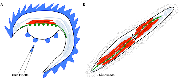
- If using the dissected preparation8,11 (Figure 1A), perform dissections in low light.
- Place animals in a dissecting dish with a silicone-coated coverslip base that is filled with a 1 mM Ca2+ extracellular solution composed of 150 mM NaCl, 5 mM KCl, 1 mM CaCl2, 4 mM MgCl2, 10 mM glucose, 5 mM sucrose, and 15 mM HEPES (pH 7.3, -340 Osm).
- Glue down animals using the liquid topical skin adhesive with blue coloring along the dorsal side of the worm and make a lateral cuticle incision along the glue/worm interface using glass needles.
- Use a mouth pipette to clear the internal viscera from the worm cavity.
- Glue down the cuticle flap of the animal to expose the ventral medial body wall muscles for imaging.
- If using the nanobead preparation (Figure 1B)
- Make a molten 5% agarose solution using ddH2O to a final volume of 100 mL.
- Using a Pasteur pipette, place a drop of molten agarose solution onto a glass slide and immediately place a second glass slide over the top, perpendicular to the first, using gentle pressure to create an even agarose pad. Remove the top slide before use.
- Add approximately 4 µL of polystyrene nanobeads (Table of Materials) to the middle of the agarose pad.
- In the low light, pick 4-6 C. elegans into the nanobead solution, making sure animals do not lay on top of each other, and carefully place a coverslip on the top.
- Place the prepared slide or dissection dish onto the microscope, and find and focus on a worm using 10x magnification and dim bright field illumination.
- Switch to 60x magnification and RCaMP fluorescence excitation to identify a ventromedial body wall muscle that is anterior to the vulva and in the correct focal plane.
NOTE: Muscles anterior to the vulva are selected as they reflect muscles commonly stimulated in electrophysiology experiments. - Change the image pathway from the eyepiece to the camera by pulling out the toggle and clicking Live within the data acquisition software (Table of Materials).
NOTE: Make sure the blue light stimulation pathway is turned off at this point to ensure that the channelrhodopsin does not get activated before capturing images. - Focus the image within the data acquisition software using the microscope fine focus.
- Once the muscle is clearly in focus, turn off the live image by unclicking the Live button.
- Click the ROI button in the data acquisition software and create a box around the muscle being focused on.
- On the stimulator, switch on the blue light stimulation pathway that has been previously programmed in step 1.10.
- Click Acquire in the imaging software to capture the image through the CMOS camera. To do this, set the exposure time to 10 s with 1,000 frames and 2x binning.
3. Data analysis
- Open the data file in the imaging software (Table of Materials) and, using the Polygon Tool, outline the muscle of interest. This is the ROI.
- Go to Image | Stacks | Plot Z-axis profile and export the resulting data points into the spreadsheet software (Table of Materials). This function plots the ROI selection mean value versus the time point.
- Move the muscle ROI created with the Polygon tool outside of the animal to get a background fluorescence measurement using the steps outlined in 3.2. Export the resulting data into the spreadsheet workbook.
- In the spreadsheet workbook, subtract the background fluorescence values from the muscle fluorescence values at each time point. This provides the background subtracted fluorescent signal.
- Average the background subtracted fluorescence for the first 2 s of data points. This will give the baseline fluorescence measurement, F.
- Use the baseline fluorescence measurement to calculate the normalized fluorescence level at each time point. To do this, use the equation (ΔF/F)+1. ΔF represents (F(t)-F), where F(t) is the fluorescence measurement at any given time point and F is the baseline value. The +1 is added as a y-axis offset.
- Repeat steps 3.2-3.6 for each muscle image that is collected. Using single or multiple muscle cells per image is at the discretion of the researcher. The n of the experiment can be determined by performing a power analysis.
- Use the processed data from steps 3.2-3.6 to make a trace of the fluorescent values in graphing software according to the manufacturer's instructions (Table of Materials).
- From this trace, measure the kinetics of the calcium transient, such as rise to peak time and half decay time, using the tools provided in the graphing software as per the manufacturer's instructions.
Representative Results
This technique evaluated changes in mutants thought to affect the calcium handling or muscle depolarization. Baseline fluorescence levels and fluorescent transients were visualized and resting cytosolic calcium and calcium kinetics within the muscle were evaluated. It is important that the animals were grown on all-trans retinal for at least three days to ensure the successful incorporation of retinal, thereby subsequently activating the channelrhodopsin (Figure 2A). If animals are not exposed to all-trans retinal, no muscle calcium transient is triggered (Figure 2B). Although these animals can still be used to evaluate baseline cytosolic calcium levels, any dynamic changes in calcium will not be captured. Additionally, as an internal control for animal or dissection health, the animal will contract following blue light stimulation. A recording with a muscle contraction that causes minimal gross movement of the worm's body was selected for data collection when using nanobeads for immobilization. If the muscle used to collect the raw fluorescence values contracts vigorously, causing the whole body of the worm to move, the transient trace will reflect this motion artifact (Figure 2C) and should be discarded from quantification.
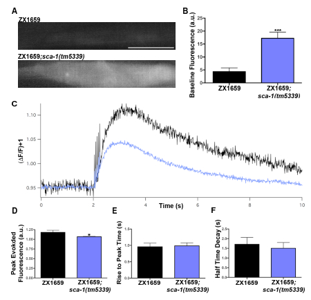
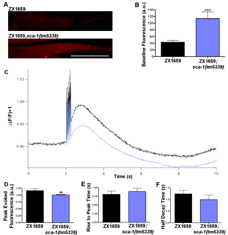
A mutation mapping to the sca-1 gene locus was isolated from a screen for mutations impacting C. elegans muscle nicotinic receptor localization. The C. elegans sca-1 gene encodes the only homolog of the sarco(endo)plasmic reticular calcium ATPase (SERCA) in the worm and is the only SERCA pump present in body wall muscles12,13. Loss-of-function sca-1 mutants were predicted to exhibit changes in the muscle calcium handling based on the important role SERCA plays in maintaining calcium homeostasis in mammalian muscles14,15,16,17,18. The GECI RCaMP was expressed in the body wall muscles and the blue light-activated channelrhodopsin was expressed in excitatory cholinergic neurons to evaluate both the intracellular baseline calcium and calcium dynamics in sca-1 mutants. With the dissected preparation technique, increases in baseline levels of RCaMP fluorescence were observed in the sca-1 mutant when compared to the control (Figure 3A,B), suggesting that loss of SERCA function leads to elevated levels of resting cytoplasmic calcium. When calcium dynamics were examined, following presynaptic stimulation triggered with a train of 5, 2 ms blue light pulses (Figure 3C), there was a significant decrease in peak calcium levels in the sca-1(tm5339) mutants as compared to the control (Figure 3D), which may reflect reduced calcium stores in the SR. However no change was seen in the rise to peak time or the half decay time (Figure 3E,F). This suggests that evoked changes in cytosolic calcium from either external source such as calcium entry through acetylcholine receptors and/or voltage-gated calcium channels as well as from internal stores are not affected in sca-1(tm5339) mutants. Similar results can be observed using the nanobead preparation, as shown in Figure 419. This recapitulation of data provides evidence that both of these techniques are physiological for measuring body wall muscle resting cytoplasmic calcium levels as well as observing stimulated calcium transients. This also demonstrates that the dissection of the worm does not cause damage to the animal that alters its calcium handling or the ability to depolarize postsynaptic muscles.
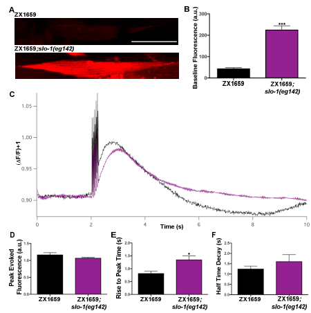
This protocol can also be used to examine mutants that indirectly impact calcium handling in C. elegans. Loss of function slo-1(eg142) mutants were evaluated to demonstrate this. The slo-1 gene encodes a calcium-activated BK potassium channel, which is expressed in both neurons and muscles20,21. Studies have previously described a role for SLO-1 in body wall muscles, demonstrating that loss of function mutants have a defect in postsynaptic repolarization following muscle action potentials20,21. Possible changes in baseline calcium levels and calcium transient dynamics due to this hyperexcitability were examined using nanobead immobilization. When baseline levels of RCaMP were measured, slo-1(eg142) mutants displayed increase fluorescence as compared to the controls (Figure 5A,B)19. This suggests that BK channels may also regulate baseline levels of cytosolic calcium. No changes in peak calcium levels were seen as compared to the control (Figure 5C,D) when evaluating the kinetics of the evoked calcium transient. The slo-1(eg142) mutants, however, exhibited a significantly increased rise to peak time as compared to the control (Figure 5E). This may reflect a previously reported increase in presynaptic excitability20,21. There was, however, no significant change in the half decay time in slo-1(eg142) mutants as compared to the control (Figure 5F). Together, these data demonstrate that this method can be used to evaluate the effects of pre- and postsynaptic mutations on both resting and stimulated body wall muscle calcium.
| Serial Setting | Value |
| Answer timeout | 500 |
| Baud rate | 57600 |
| DelayBetweenCharsMs | 0 |
| Handshaking | Off |
| Parity | None |
| StopBits | 1 |
| Verbose | 0 |
Table 1: Serial port settings for microcontroller plugin, which controls an external microcontroller board. This is used to program external timing pulses to control fluorescence excitation.

Figure 1: Graphical representation of different immobilization techniques. (A) This graphic demonstrates the dissection technique for immobilization. The body of the worm is glued down (blue) around the dorsal side of the animal. A dorsal incision along the cuticle generates a cuticle flap. This flap has been glued down to expose the intact NMJs formed between synapses of the ventral nerve cord in green (channelrhodopsin) and RCaMP expressing muscles in red. (B) This graphic demonstrates the nanobead immobilization technique. The intact worm is surrounded by a representation of the nanobeads in solution, that when compressed by the overlying coverslip, immobilize the worm. Due to the transparency of C. elegans, the ventral nerve cord can be visualized in green (channelrhodopsin) and RCaMP expressing muscles can be visualized in red. The ovoid in the middle of the worm represents the vulva, which is a landmark identifying the ventral body wall muscles. Please click here to view a larger version of this figure.

Figure 2: Representative traces of calcium transients. (A) Single trace representation of a calcium transient evoked by a train of 5, 2 ms blue light pulses at 50 ms interpulse intervals, recorded in an animal with limited body movement (- movement) grown in the presence of all-trans retinal (+ all-trans retinal). The spike artifacts are the blue light stimulation pulses, as both LEDs are co-illuminated simultaneously. (B) Single trace representation showing the absence of a calcium transient in response to a train of 5, 2 ms blue light pulses with 50 ms interpulse intervals in an animal that has not been exposed to all-trans retinal (- all-trans retinal, – movement). (C) Single trace representation of a calcium transient that has been evoked by a train of 5, 2 ms blue light pulses with 50 ms interpulse intervals, recorded in an animal that had significant muscle contraction leading to movement artifacts (+ movement, + all-trans retinal). Please click here to view a larger version of this figure.

Figure 3: Baseline calcium levels and evoked calcium transients from control and sca-1(tm5339) samples prepared using the dissection technique. (A) Representative images of baseline RCaMP fluorescence in sca-1(tm5339) mutants and controls. Scale bar = 50 µm. (B) Baseline fluorescence levels quantification in sca-1(tm5339) mutants (n=13) and control (n=9). (C) Ensemble average traces of evoked calcium transients for sca-1(tm5339) mutants and control animals. (D) Quantification of peak RCaMP fluorescence from channelrhodopsin evoked calcium responses in sca-1(tm5339) mutants (n=12) and controls (n=9). (E) Quantification of the rise to peak time of channelrhodopsin evoked calcium transients in sca-1(tm5339) mutants (n=12) and controls (n=8). (F) Half decay time quantification of channelrhodopsin evoked calcium transients in sca-1(tm5339) mutants (n=12) and controls (n=7). Statistically significant values were: not significant p>0.05, * p≤0.05, ** p≤0.01, *** p≤0.001. Error bars represent mean ± SEM. Data were normalized using Shapiro-Wilk tests and significance was determined with a Mann-Whitney test for non-normal distributions. Please click here to view a larger version of this figure.

Figure 4: Baseline calcium levels and evoked calcium transients from control and sca-1(tm5339) samples prepared using nanobead immobilization. (A) Representative images of baseline fluorescence in sca-1(tm5339) mutants and controls, scale bar 50 µm. (B) Quantification of baseline RCaMP fluorescence levels in sca-1(tm5339) mutants (n=14) and the control (n=13). (C) Average of evoked calcium transients for sca-1(tm5339) mutants and controls. (D) Quantification of peak RCaMP fluorescence following evoked calcium response in sca-1(tm5339) mutants (n=14) and controls (n=13). (E) Quantification of rise to peak time of evoked calcium transients in sca-1(tm5339) mutants (n=14) and controls (n=12). (F) Quantification of half decay time of evoked calcium transients in sca-1(tm5339) mutants (n=14) and control (n=13). Figure is adapted from19. Statistically significant values were: not significant p > 0.05, * p ≤ 0.05, ** p ≤ 0.01, *** p ≤ 0.001. Error bars show mean ± SEM. Data normality was assessed using Shapiro-Wilk tests and significance was determined with a Mann-Whitney test for non-normal distributions. This figure has been modified with permission from19. Please click here to view a larger version of this figure.

Figure 5: Baseline calcium levels and evoked calcium transients from control and slo-1(eg142) samples prepared using nanobeads immobilization. (A) Representative images of baseline fluorescence in slo-1(eg142) mutants and controls, scale bar 50 µm. (B) Quantification of baseline RCaMP fluorescence levels in slo-1(eg142) mutants (n=29) and the control (n=13). (C) Average of evoked calcium transients for slo-1(eg142) mutants and controls. (D) Quantification of peak RCaMP fluorescence following evoked calcium response in slo-1(eg142) mutants (n=29) and controls (n=13). (E) Quantification of rise to peak time of evoked calcium transients in slo-1(eg142) mutants (n=29) and controls (n=13). (F) Quantification of half decay time of evoked calcium transients in slo-1(eg142) mutants (n=11) and controls (n=13). Figure is adapted from19. Statistically significant values were: not significant p > 0.05, * p ≤ 0.05, ** p ≤ 0.01, *** p ≤ 0.001. Error bars show mean ± SEM. Data normality was assessed using Shapiro-Wilk tests and significance was determined with a Mann-Whitney test for non-normal distributions. This figure has been modified with permission from19. Please click here to view a larger version of this figure.
Discussion
GECIs are a powerful tool in C. elegans neurobiology. Previous work has utilized calcium imaging techniques to examine a wide variety of functions in both neurons and muscle cells, including sensory and behavioral responses, with varied methods of stimulation. Some studies have used chemical stimuli to trigger calcium transients in sensory ASH neurons22,23 or to induce calcium waves in pharyngeal muscles24. Another group utilized mechanical stimulation while worms were held in microfluidic chips to examine calcium responses in touch receptor neurons25. Still, others have employed electrical stimulation to visualize calcium changes in both neurons26 and body wall muscles27. What is common between these studies is the requirement of external stimuli, such as solutions or equipment, to trigger calcium transients. The protocol outlined here capitalizes on the control gained from optogenetic stimulation, which provides neuronal specificity, coupled with the functional analysis of muscle expressed GECIs2 in the same experimental paradigm.
Two forms of worm immobilization are described and, while each method should be evaluated when addressing experimental needs, both dissected and physiologically intact preparations produce complementary results allowing investigators to use either approach in their research. There is an increased technical challenge to using the dissection technique, with the user being required to master both precise use of the surgical glue and microdissection without damaging the nerve cord or muscles. Additionally, due to the level of difficulty of the dissection, only gravid adults should be used, which could limit the experimental time points. Furthermore, the dissection technique is also much harder to perform on mutants that produce small, thin, or sickly animals, again possibly limiting experimental parameters. The advantage of this technique is that it limits the body motion artifact resulting from depolarization, gluing prevents excess movement of the worm's body, and also provides access to the NMJ for solution changes and drug applications. Additionally, each preparation exposes the same area of the worm, which guarantees body wall muscles will be consistently in focus. The second technique, using nanobeads to immobilize the worms, is less involved and can be quickly learned. Also, any age animals can be used with this method, allowing for changes in calcium handling through development to be monitored. Care does need to be taken when placing the worms into the nanobead solution, as too much manipulation of the animals may cause them to burst. Also, the coverslip must not be moved once placed on top of the animals, as this may cause the animals to roll or twist on themselves, again leading to the damage. Although using the nanobeads can provide a high-throughput assay in which the animals could potentially be recovered, there is no standardization of the animal's body position, thus every animal may not have body wall muscles in a clear focal plane. If the experiment calls for a large number of animals, different methods of immobilization may be necessary, such as agar-based micro-wells, which allow for the bulk calcium fluorescence imaging28. Regardless of the limitations of these two techniques, either can be utilized to obtain reliable, easily quantifiable calcium transients within C. elegans body wall muscles.
C. elegans provide many advantages as an in vivo system for the functional imaging studies. The animals are transparent, allowing the pairing of optogenetic neuronal depolarization through the use of channelrhodopsin with the GECI RCaMP to measure the resulting postsynaptic calcium transients in the muscle. Pre-stimulation levels of RCaMP fluorescence can also be used to assess baseline cytosolic calcium levels. The genetic tractability of C. elegans makes the study of novel mutations that may affect calcium homeostasis or handling easy to pursue, often with readily available mutant alleles. Using the techniques outlined in this protocol, postsynaptic calcium-imaging data can be obtained efficiently and at relatively low cost, making this an attractive system for a wide range of imaging experiments.
Divulgazioni
The authors have nothing to disclose.
Acknowledgements
The authors thank Dr. Alexander Gottschalk for ZX1659, the RCaMP and channelrhodopsin containing worm strain, Dr. Hongkyun Kim for the slo-1(eg142) worm strain, and the National Bioresource Project for the sca-1(tm5339) worm strain.
Materials
| all-trans retinal | Sigma-Aldrich | R2500 | Necessary for excitation of channel rhodopsin |
| Amber LED | RCaMP illumination | ||
| Arduino UNO | Mouser | 782-A000066 | Controls fluorescence illumination |
| Blue LED | channelrhodopsin illumination | ||
| BX51WI microscope | Olympus | Fixed state compound microscope | |
| Current controlled low noise linear power supply | Ametek | Sorenson | Controls LED intensity |
| Igor Pro | Wavemetrics | Wavemetrics.com | Graphing software |
| ImageJ | NIH | imagej.nih.gov | Image processing software |
| LUMFLN 60x water NA 1.4 | Olympus | Water immersion objective for dissected preparation | |
| Master-8 Stimulator | A.M.P.I | Master timer for image acquisition and LED illumination | |
| Micro-Manager | micro-manager.org | Controls camera acquisition and LED excitiation | |
| Microsoft Excel | Microsoft | Spreadsheet software | |
| pco.edge 4.2 CMOS camera | pco. | 4.2 | High-speed camera |
| PlanApo N 60x oil NA 1.4 | Olympus | Oil immersion objective for nanobead preparation | |
| Polybead microspheres | Polysciences, Inc. | 00876-15 | For worm immobilization |
| solid state switches | Sensata Technologies | Crydom CMX100D6 | Controls timing of LED illumination |
| Transgenic strain, sca-1(tm5339); [zxIs6{Punc17::chop-2 (h134R)::yfp,lin-15(+)}; Pmyo3::RCaMP35] |
Richmond Lab | SY1627 | Excitatory neuronal channelrhodopsin and body wall muscle RCaMP expressing worm line with SERCA mutant allele |
| Transgenic strain, slo-1 (eg142); [zxIs6{Punc17::chop-2 (h134R)::yfp,lin-15(+)}; Pmyo3::RCaMP35] |
Richmond Lab | Excitatory neuronal channelrhodopsin and body wall muscle RCaMP expressing worm line with calcium-activated BK potassium channel mutant allele | |
| Transgeneic strain, [zxIs6{Punc17::chop-2 (h134R)::yfp,lin-15(+)}; Pmyo3::RCaMP35] |
Gottschalk Lab | ZX1659 | Excitatory neuronal channelrhodopsin and body wall muscle RCaMP expressing worm line |
Riferimenti
- Nakai, J., Ohkura, M., Imoto, K. A high signal-to-noise Ca2+ probe composed of a single green fluorescent protein. Nature Biotechnology. 19 (2), 137-141 (2001).
- Akerboom, J., et al. Genetically encoded calcium indicators for multi-color neural activity imaging and combination with optogenetics. Frontiers in Molecular Neuroscience. 6, 1-29 (2013).
- Nagel, G., et al. Light Activation of Channelrhodopsin-2 in Excitable Cells of Caenorhabditis elegans Triggers Rapid Behavioral Responses. Current Biology. 15 (24), 2279-2284 (2005).
- Liewald, J. F., et al. Optogenetic analysis of synaptic function. Nature Methods. 5 (10), 895-902 (2008).
- Fang-Yen, C., Gabel, C. V., Samuel, A. D. T., Bargmann, C. I., Avery, L. Laser Microsurgery in Caenorhabditis elegans. Methods Cell Biology. 107, 177-206 (2012).
- Lewis, J. A., et al. Cholinergic Receptor Mutants of the Nematode Caenorhabditis elegans. The Journal of Neuroscience. 7 (10), 3059-3071 (1987).
- Lewis, J. A., Wu, C. H., Berg, H., Levine, J. H. The genetics of levamisole resistance in the nematode Caenorhabditis elegans. Genetica. 95 (4), 905-928 (1980).
- Richmond, J. E., Jorgensen, E. M. One GABA and two acetylcholine receptors function at the C. elegans neuromuscular junction. Nature Neuroscience. 9, 791-798 (1999).
- Kim, E., Sun, L., Gabel, C. V., Fang-Yen, C. Long-Term Imaging of Caenorhabditis elegans Using Nanoparticle-Mediated Immobilization. PLoS ONE. 8 (1), 1-6 (2013).
- Edelstein, A., Amodaj, N., Hoover, K., Vale, R., Stuurman, N. Computer control of microscopes using umanager. Current Protocols in Molecular Biology. (Suppl 92), 1-17 (2010).
- Richmond, J. Dissecting and Recording from The C. Elegans Neuromuscular Junction. Journal of Visualized Experiments. (24), 1-4 (2009).
- Hoon Cho, J., Bandyopadhyay, J., Lee, J., Park, C. S., Ahnn, J. Two isoforms of sarco/endoplasmic reticulum calcium ATPase (SERCA) are essential in Caenorhabditis elegans. Gene. 261, 211-219 (2000).
- Zwaal, R. R., et al. The Sarco-Endoplasmic Reticulum Ca2+ ATPase Is Required for Development and Muscle Function in Caenorhabditis elegans. The Journal of Biological Chemistry. 276 (47), 43557-43563 (2001).
- Stammers, A. N., et al. The regulation of sarco(endo)plasmic reticulum calcium-ATPases (SERCA). Canadian Journal of Physiology and Pharmacology. 93, 1-12 (2015).
- Clapham, D. E. Calcium Signaling. Cell. 131, 1047-1058 (2007).
- Hovnanian, A. Serca pumps and human diseases. Calcium Signalling and Disease: Molecular Pathology of Calcium. , 337-363 (2007).
- Periasamy, M., Kalyanasundaram, A. SERCA pump isoforms: Their role in calcium transport and disease. Muscle and Nerve. 35 (4), 430-442 (2007).
- Gehlert, S., Bloch, W., Suhr, F. Ca2+-Dependent Regulations and Signaling in Skeletal Muscle: From Electro-Mechanical Coupling to Adaptation. International Journal of Molecular Sciences. 16, 1066-1095 (2015).
- Martin, A. A., Richmond, J. E. The sarco(endo)plasmic reticulum calcium ATPase SCA-1 regulates the Caenorhabditis elegans nicotinic acetylcholine receptor ACR-16. Cell Calcium. 72, 104-115 (2018).
- Wang, Z., Saifee, O., Nonet, M. L., Salkoff, L. SLO-1 Potassium Channels Control Quantal Content of Neurotransmitter Release at the C. elegans Neuromuscular Junction. Neuron. 32, 867-881 (2001).
- Abraham, L. S., Oh, H. J., Sancar, F., Richmond, J. E., Kim, H. An Alpha-Catulin Homologue Controls Neuromuscular Function through Localization of the Dystrophin Complex and BK Channels in Caenorhabditis elegans. PLoS Genetics. 6 (8), 1-13 (2010).
- Gourgou, E., Chronis, N. Chemically induced oxidative stress affects ASH neuronal function and behavior in C. elegans. Scientific Reports. 6, 1-9 (2016).
- Zahratka, J. A., Williams, P. D. E., Summers, P. J., Komuniecki, R. W., Bamber, B. A. Serotonin differentially modulates Ca2+ transients and depolarization in a C. elegans nociceptor. Journal of Neurophysiology. 113 (4), 1041-1050 (2015).
- Kerr, R., et al. Optical imaging of calcium transients in neurons and pharyngeal muscle of C. elegans. Neuron. 26 (3), 583-594 (2000).
- Nekimken, A. L., et al. Pneumatic stimulation of C. elegans mechanoreceptor neurons in a microfluidic trap. Lab Chip. 17 (6), 1116-1127 (2017).
- Chung, S. H., Sun, L., Gabel, C. V. In vivo Neuronal Calcium Imaging in C. elegans. Journal of Visualized Experiments. (74), 1-9 (2013).
- Jospin, M., Jacquemond, V., Mariol, M. C., Ségalat, L., Allard, B. The L-type voltage-dependent Ca2+channel EGL-19 controls body wall muscle function in Caenorhabditis elegans. Journal of Cell Biology. 159 (2), 337-347 (2002).
- Wabnig, S., Liewald, J. F., Yu, S., Gottschalk, A. High-Throughput All-Optical Analysis of Synaptic Transmission and Synaptic Vesicle Recycling in Caenorhabditis elegans. PLoS ONE. , 1-26 (2015).

