JoVE Science Education
Microbiology
A subscription to JoVE is required to view this content.  Sign in or start your free trial.
JoVE Science Education Microbiology
16S rRNA Sequencing: A PCR-based Technique to Identify Bacterial Species
  • 00:01Concepts
  • 03:13gDNA Isolation and Quality Check
  • 03:43Isolation of gDNA and gDNA Quality Check
  • 05:52Amplification and Purification of 16S rRNA Gene by PCR
  • 07:20Analysis of the DNA Sequences
  • 09:02Sequence Assembly and Database Search

Secuenciación del ARNr 16s: Una técnica basada en PCR para identificar especies bacterianas

English

Share

Overview

Fuente: Ewa Bukowska-Faniband1, Tilde Andersson1, Rolf Lood1
1 Departamento de Ciencias Clínicas Lund, División de Medicina de Infecciones, Centro Biomédico, Universidad de Lund, 221 00 Lund, Suecia

El planeta Tierra es un hábitat para millones de especies bacterianas, cada una de las cuales tiene características específicas. La identificación de especies bacterianas se utiliza ampliamente en la ecología microbiana para determinar la biodiversidad de muestras ambientales y microbiología médica para diagnosticar pacientes infectados. Las bacterias se pueden clasificar utilizando métodos de microbiología convencionales, como la microscopía, el crecimiento en medios específicos, las pruebas bioquímicas y serológicas y los ensayos de sensibilidad a los antibióticos. En las últimas décadas, los métodos de microbiología molecular han revolucionado la identificación bacteriana. Un método popular es la secuenciación del gen del ARN ribosomal 16S (ARN RRNA). Este método no sólo es más rápido y preciso que los métodos convencionales, sino que también permite la identificación de cepas que son difíciles de cultivar en condiciones de laboratorio. Además, la diferenciación de las cepas a nivel molecular permite la discriminación entre bacterias fenotípicamente idénticas (1-4).

El ARNR 16S se une a un complejo de 19 proteínas para formar una subunidad 30S del ribosoma bacteriano (5). Está codificado por el gen 16S rRNA, que está presente y altamente conservado en todas las bacterias debido a su función esencial en el ensamblaje ribosoma; sin embargo, también contiene regiones variables que pueden servir como huellas dactilares para determinadas especies. Estas características han hecho del gen rRNA 16S un fragmento genético ideal para ser utilizado en la identificación, comparación y clasificación filogenética de bacterias (6).

La secuenciación del gen rRNA 16S se basa en la reacción en cadena de la polimerasa (PCR) (7-8) seguida de la secuenciación del ADN (9). La PCR es un método de biología molecular utilizado para amplificar fragmentos específicos de ADN a través de una serie de ciclos que incluyen:

i) Desnaturalización de una plantilla de ADN de doble cadena
ii) Recocido de imprimaciones (oligonucleótidos cortos) que son complementarios a la plantilla
iii) Extensión de imprimaciones por la enzima ADN polimerasa, que sintetiza una nueva cadena de ADN

En la Figura 1se muestra una descripción esquemática del método.

Figure 1
Figura 1: Descripción esquemática de la reacción PCR. Haga clic aquí para ver una versión más grande de esta figura.

Hay varios factores que son importantes para una reacción de PCR exitosa, uno de los cuales es la calidad de la plantilla de ADN. El aislamiento del ADN cromosómico de las bacterias se puede realizar utilizando protocolos estándar o kits comerciales. Se debe tener especial cuidado para obtener ADN que esté libre de contaminantes que puedan inhibir la reacción de PCR.

Las regiones conservadas del gen 16S rRNA permiten el diseño de pares de imprimación universales (uno hacia adelante y otro inverso) que pueden unirse y amplificar la región objetivo en cualquier especie bacteriana. La región de destino puede variar en tamaño. Mientras que algunos pares de imprimación pueden amplificar la mayor parte del gen rRNA 16S, otros amplifican sólo partes de él. En la Tabla 1 se muestran ejemplos de imprimaciones de uso común y sus sitios de enlace se muestran en la Figura 2.

Primer nombre Secuencia (5′-3′) Adelante/retroceso Referencia
8F b) AGAGTTTGATCCTGGCTCAG Adelante -1
27F AGAGTTTGATCMTGGCTCAG Adelante -10
515F GTGCCAGCMGCCGCGGTAA Adelante -11
911R GCCCCCGTCAATTCMTTTGA Marcha atrás -12
1391R GACGGGCGGTGTARCA Marcha atrás -11
1492R GGTTACCTTGTTACGACTT Marcha atrás -11

Tabla 1: Ejemplos de oligonucleótidos estándar utilizados en la amplificación de genes 16S rRNA a).
a) Las longitudes esperadas del producto PCR generado utilizando las diferentes combinaciones de imprimación se pueden estimar calculando la distancia entre los sitios de unión para la imprimación delantera y la inversa (véase la figura 2), por ejemplo, el tamaño del PCR producto con el par de imprimación 8F-1492R es de 1500 bp, y para el par de imprimación 27F-911R a 900 bp.
b) también conocido como fD1

Figure 2
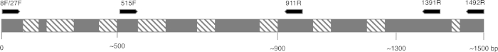
Figura 2: Figura representativa de la secuencia de rRNA 16S y los sitios de enlace de imprimación. Las regiones conservadas se colorean en gris y las regiones variables se rellenan con líneas diagonales. Para permitir la resolución más alta, la imprimación 8F y 1492R (nombre basado en la ubicación en la secuencia rRNA) se utilizan para amplificar toda la secuencia, permitiendo la secuencia de varias regiones variables del gen. Haga clic aquí para ver una versión más grande de esta figura.

Las condiciones de ciclismo para pcR(es decir, la temperatura y el tiempo necesarios para que el ADN sea desnaturalizado, recocido con imprimadores y sintetizado) dependen del tipo de polimerasa que se utiliza y de las propiedades de las imprimaciones. Se recomienda seguir las pautas del fabricante para una polimerasa en particular.

Una vez completado el programa PCR, los productos son analizados por electroforesis de gel de agarosa. Un PCR exitoso produce una sola banda de tamaño esperado. El producto debe purificarse antes de la secuenciación para eliminar las imprimaciones residuales, los desoxirribonucleótidos, la polimerasa y el tampón que estaban presentes en la reacción de PCR. Los fragmentos de ADN purificados generalmente se envían para la secuenciación a servicios de secuenciación comercial; sin embargo, algunas instituciones realizan la secuenciación del ADN en sus propias instalaciones principales.

La secuencia de ADN se genera automáticamente a partir de un cromatograma de ADN por una computadora y debe ser cuidadosamente revisada para la calidad, ya que a veces se necesita la edición manual. Siguiendo este paso, la secuencia genética se compara con las secuencias depositadas en la base de datos 16S rRNA. Se identifican las regiones de similitud y se entregan las secuencias más similares.

Procedure

1. Configurar Mientras se manipulan microorganismos, se requiere seguir buenas prácticas microbiológicas. Todos los microorganismos, especialmente las muestras desconocidas, deben tratarse como patógenos potenciales. Siga la técnica aséptica para evitar contaminar las muestras, los investigadores o el laboratorio. Lávese las manos antes y después de manipular las bacterias, use guantes y use ropa protectora. Llevar a cabo una evaluación de riesgos para el protocolo experimenta…

Applications and Summary

Identifying bacterial species is important for different researchers, as well as for those in healthcare. 16S rRNA sequencing was initially used by researchers to determine phylogenetic relationships between bacteria. In time, it has been implemented in metagenomic studies to determine biodiversity of environmental samples and in clinical laboratories as a method to identify potential pathogens. It enables a quick and accurate identification of bacteria present in clinical samples, facilitating earlier diagnosis and faster treatment of patients.

References

  1. Weisburg, W.G., Barns, S.M., Pelletier, D.A. and Lane D.J. 16S ribosomal DNA amplification for phylogenetic study. J Bacteriol. 173 (2): 697-703. (1991)
  2. Drancourt, M., Bollet, C., Carlioz, A., Martelin, R., Gayral, J.P., Raoult D. 16S ribosomal DNA sequence analysis of a large collection of environmental and clinical unidentifiable bacterial isolates. J Clin Microbiol. 38 (10):3623-3630. (2000)
  3. Woo, P.C., Lau, S.K., Teng, J.L., Tse, H., Yuen, K.Y. Then and now: use of 16S rDNA gene sequencing for bacterial identification and discovery of novel bacteria in clinical microbiology laboratories. Clin Microbiol Infect. 14 (10):908-934. (2008)
  4. Tang, Y.W., Ellis, N.M., Hopkins, M.K., Smith, D.H., Dodge, D.E., Persing, D.H. Comparison of phenotypic and genotypic techniques for identification of unusual aerobic pathogenic gram-negative bacilli. J Clin Microbiol. 36 (12):3674-3679. (1998)
  5. Tsiboli, P., Herfurth, E., Choli, T. Purification and characterization of the 30S ribosomal proteins from the bacterium Thermus thermophilus. Eur J Biochem. 226 (1):169-177. (1994)
  6. Woese, C.R. Bacterial evolution. Microbiol Rev. 51 (2):221-271. (1987)
  7. Bartlett, J.M., Stirling, D. A short history of the polymerase chain reaction. Methods Mol Biol. 226:3-6. (2003)
  8. Wilson, K.H., Blitchington, R.B., Greene, R.C. Amplification of bacterial 16S ribosomal DNA with polymerase chain reaction. J Clin Microbiol. 28 (9):1942-1946. (1990)
  9. Shendure, J., Balasubramanian, S., Church, G.M., Gilbert, W., Rogers, J., Schloss, J.A., Waterston, R.H. (2017) DNA sequencing at 40: past, present and future. Nature. 550:345-353.
  10. Lane, D.J. 16S/23S rRNA sequencing. (1991) In Nucleic acid techniques in bacterial systematics. (Goodfellow, M. and Stackebrandt, E., eds.) p.115-175. Wiley and Sons, Chichester, United Kingdom.
  11. Turner, S., Pryer, K.M., Miao, V.P., Palmer, J.D. (1999) Investigating deep phylogenetic relationships among cyanobacteria and plastids by small subunit rRNA sequence analysis. J Eukaryot Microbiol. 46:327-338.
  12. Fredricks, D.N., Relman, D.A. (1998) Improved amplification of microbial DNA from blood cultures by removal of the PCR inhibitor sodium polyanetholesulfonate. J Clin Microbiol. 36:2810-2816.
  13. Wilson, K. Preparation of genomic DNA from bacteria. (2001) Curr Protoc Mol Biol. Chapter 2:Unit 2.4.
  14. Wright, M. H., Adelskov, J., Greene, A.C. (2017) Bacterial DNA extraction using individual enzymes and phenol/chloroform separation. J Microbiol Biol Educ. 18:18.2.48.
  15. Huang, X., Madan, A. (1999). CAP3: A DNA sequence assembly program. Genome Res. 9:868-877.

Transcript

Earth is home to millions of bacterial species, each with unique characteristics. Identifying these species is critical in evaluating environmental samples. Doctors also need to distinguish different bacterial species to diagnose infected patients.

To identify bacteria, a variety of techniques can be employed, including microscopic observation of morphology or growth on a specific media to observe colony morphology. Genetic analysis, another technique for identifying bacteria has grown in popularity in recent years, due in part to 16S ribosomal RNA gene sequencing.

The bacterial ribosome is a protein RNA complex consisting of two subunits. The 30S subunit, the smaller of these two subunits, contains 16S rRNA, which is encoded by the 16S rRNA gene contained within the genomic DNA. Specific regions of 16S rRNA are highly conserved, due to their essential function in ribosome assembly. While other regions, less critical to function, may vary among bacterial species. The variable regions in 16S rRNA, can serve as unique molecular fingerprints for bacterial species, allowing us to distinguish phenotypically identical strains.

After obtaining a quality sample of gDNA, PCR of the 16S rRNA encoding gene can begin. PCR is a commonly used molecular biology method, consisting of cycles of denaturation of the double-stranded DNA template, annealing of universal primer pairs, which amplify highly conserved regions of the gene, and the extension of primers by DNA polymerase. While some primers amplify most of the 16S rRNA encoding gene, others only amplify fragments of it. After PCR, the products can be analyzed via agarose gel electrophoresis. If amplification was successful, the gel should contain a single band of an expected size, depending upon the primer pair used, up to 1500bp, the approximate length of the 16S rRNA gene.

After purification and sequencing, the obtained sequences can then be entered into the BLAST database, where they can be compared with reference 16S rRNA sequences. As this database returns matches based on the highest similarity, this allows confirmation of the identity of the bacteria of interest. In this video, you will observe 16S rRNA gene sequencing, including PCR, DNA sequence analysis and editing, sequence assembly and database searching.

When handling microorganisms, it is essential to follow good microbiological practice, including using aseptic technique and wearing appropriate personal protective equipment. After performing an appropriate risk assessment for the microorganism or environmental sample of interest, obtain a test culture. In this example, a pure culture of Bacillus subtilis is used.

To begin, grow your microorganism on a suitable medium in the appropriate conditions. In this example, Bacillus subtilis 168 is grown in LB broth overnight in a shaking incubator set to 200 rpm at 37 degrees Celsius. Next, use a commercially available kit to isolate genomic DNA or gDNA from 1.5 milliliters of the B. subtilis overnight culture.

To check the quality of the isolated DNA, first mix five microliters of the isolated gDNA with one microliter of DNA gel loading dye. Then, load the sample onto a 0.8% agarose gel, containing DNA staining reagent, such as SYBR safe or EtBr. After this, load a one kilobase molecular mass standard onto the gel, and run the electrophoresis until the front dye is approximately 0.5 centimeters from the bottom of the gel. Once the gel electrophoresis is complete, visualize the gel on a blue light transilluminator. The gDNA should appear as a thick band, above 10 kilobase in size and have minimal smearing.

After this, to create serial dilutions of the gDNA, label three microcentrifuge tubes as 10X, 100X, and 1000X. Then, use a pipette to dispense 90 microliters of sterile distilled water into each of the tubes. Next, add 10 microliters of the gDNA solution to the 10X tube. Pipette the whole volume up and down to ensure the solution is mixed thoroughly. Then, remove 10 microliters of the solution from the 10X tube and transfer this to the 100X tube. Mix the solution as previously described. Finally, transfer 10 microliters of the solution in the 100X tube, to the 1000X tube.

To begin the PCR protocol, thaw the necessary reagents on ice. Then, prepare the PCR master mix. Since the DNA polymerase is active at room temperature, the reaction set up must occur on ice. Aliquot 49 microliters of the master mix into each of the PCR tubes. Then, add one microliter of template to each of the experimental tubes and one microliter of sterile water to the negative control tube, pipetting up and down to mix. After this, set the PCR machine according to the program described in the table. Place the tubes into the thermocycler and start the program.

Once the program is complete, examine the quality of your product via agarose gel electrophoresis, as previously demonstrated. A successful reaction using the described protocol should yield a single band of approximately 1.5 kilobase. In this example, the sample containing 100X diluted gDNA yielded the highest quality product. Next, purify the best PCR product, in this case, the 100X gDNA, with a commercially available kit. Now the PCR product can be sent for sequencing.

In this example, the PCR product is sequenced using forward and reverse primers. Thus, two data sets, each containing a DNA sequence and a DNA chromatogram, are generated: one for the forward primer and the other for the reverse primer. First, examine the chromatograms generated from each primer. An ideal chromatogram should have evenly spaced peaks with little to no background signals.

If the chromatograms display double peaks, multiple DNA templates may have been present in the PCR products and the sequence should be discarded. If the chromatograms contained peaks of different colors in the same location, the sequencing software likely miscalled nucleotides. This error can be manually identified and corrected in the text file. The presence of broad peaks in the chromatogram indicates a loss of resolution, which causes miscounting of the nucleotides in the associated regions. This error is difficult to correct and mismatches in any of the subsequent steps should be treated as unreliable. Poor chromatogram reading quality, indicated by the presence of multiple peaks, usually occurs at the five prime and three prime ends of the sequence. Some sequencing programs remove these low quality sections automatically. If your sequence was not truncated automatically, identify the low quality fragments and remove their respective bases from the text file.

Use a DNA assembly program to assemble the two primer sequences into one continuous sequence. Remember, sequences obtained using forward and reverse primers should partially overlap. In the DNA assembly program, insert the two sequences in FASTA format into the appropriate box. Then, click the submit button and wait for the program to return the results.

To view the assembled sequence, click on Contigs in the results tab. Then, to view the details of the alignment, select assembly details. Navigate to the website for the basic local alignment search tool, or BLAST, and select the nucleotide BLAST tool to compare your sequence to the database. Enter your sequence into the query sequence text box and select the appropriate database in the scroll down menu. Finally, click the BLAST button on the bottom of the page, and wait for the tool to return the most similar sequences from the database.

In this example, the top hit is B. subtilis strain 168, showing 100% identity with the sequence in the BLAST database. If the top hit does not show 100% identity to your expected species or strain, click on the sequence which most closely matches your query to see the details of the alignment. Aligned nucleotides will be joined by short vertical lines and mismatched nucleotides will have gaps between them. Focusing on the identified mismatched regions, revise the sequence and repeat the BLAST search if desired.

Tags

Cite This
JoVE Science Education Database. JoVE Science Education. 16S rRNA Sequencing: A PCR-based Technique to Identify Bacterial Species. JoVE, Cambridge, MA, (2023).