Measuring Volatile and Non-volatile Antifungal Activity of Biocontrol Products
Özet
We describe a modified agar-based method designed to quantify the antifungal effects of plant-derived products. Both volatile and non-volatile contributions to the antifungal activity can be assessed through this protocol. In addition, efficacy against fungi can be measured at key developmental stages in a single experimental setup.
Abstract
The protocol described is based on a plug-transfer technique that allows accurate determination of microorganism quantities and their developmental stages. A specified number of spores are spread on an agar plate. This agar plate is incubated for a defined period to allow the fungi to reach the expected developmental stage, except for spores where incubation is not required. Agar plugs covered by spores, hyphae, or mycelium are next withdrawn and transferred onto agar media containing the antifungal compound to be tested either placed at a distance from the fungi or in contact. This method is applicable to test both liquid extracts and solid samples (powders). It is particularly well suited for quantifying the relative contributions of volatile and non-volatile agents in bioactive mixtures and for determining their effects, specifically on spores, early hyphae, and mycelium.
The method is highly relevant for the characterization of the antifungal activity of biocontrol products, notably plant-derived products. Indeed, for plant treatment, the results can be used to guide the choice of mode of application and to establish the trigger thresholds.
Introduction
Global losses of fruits and vegetables can reach up to 50% of production1 and result mostly from food decay caused by fungi spoilage in field or during post-harvest storage2,3, despite the extensive employment of synthetic fungicides since the middle of the twentieth century4. The use of these substances is being reconsidered since it represents serious environmental and health hazards. As the harmful consequences of their use are showing up throughout ecosystems and evidence of potential health impacts has accumulated5,6, novel alternatives to old prophylactic strategies are being developed for pre- and post-harvest treatments7,8,9. Hence the challenge we face is two-fold. Novel fungicidal strategies must, firstly, maintain the levels of efficacy of food protection against phytopathogens and concomitantly, secondly, contribute to dramatically reducing the environmental footprint of agricultural practices. To fulfill this ambitious goal, strategies inspired by the natural defenses evolved in plants are being proposed as more than 1000 plants species have been highlighted for their antimicrobial properties8. For instance, plants which have developed natural fungicides to fight phytopathogens are a novel resource in exploring the development of new biocontrol products2. Essential oils are flagship molecules of this type. For example, Origanum essential oil protects tomato plants against gray mold in greenhouses 10 and Solidago canadensis L. and cassia essential oils have been shown to preserve post-harvested strawberries from gray mold damage11,12. These examples illustrate that biocontrol and notably plant-derived products represent a solution that combines biological efficacy and environmental sustainability.
Thus, plants are an important resource of molecules of potential interest for the crop-protection industry. However only a handful of plant products have been proposed to be used as biocontrol products even though they are generally recognized as safe, non-phytotoxic and eco-friendly2. Some difficulties in the transposition from the lab to the field have been observed, such as efficacy decreasing once applied in vivo2,9. Thus, it becomes important to improve the ability of lab tests to better predict field efficacy. In this context, antifungal testing methods for plant-derived products are necessary both to evaluate their antifungal efficacy and to define their optimal conditions for use. Specifically, biocontrol products are generally less efficient than chemical fungicides, so a better understanding of their mode of action is important for proposing suitable formulations, to identify the mode of application in fields, and to define which developmental stage of the pathogen is vulnerable to the candidate bioproduct.
Current approaches addressing antibacterial and antifungal activities include diffusion methods such as agar-disk diffusion, dilution, bioautography and flow cytometry13. Most of these techniques, and more specifically, the standard antifungal susceptibility testing – agar-disk diffusion and dilution assays – are well-adapted for evaluating the antimicrobial activity of soluble compounds on bacterial and fungal spores in liquid suspensions14. However, these methods are generally not suitable for testing solid compounds such as dried plant powder or to quantify antifungal activity during mycelium growth as they require spore dilution or spore spreading on agar plates and/or dilution of antifungal compounds13. In the food-poisoned method, agar plates containing the antifungal agent are inoculated with a 5-7 mm diameter disk sampled from a 7-day old fungi culture without considering the precise quantity of starting mycelium. After incubation, the antifungal activity is determined as a percent of radial-growth inhibition17,18,19. With this approach we can evaluate the antifungal activity on mycelial growth. By contrast, the agar-dilution method is performed to determine the antifungal activity on spores directly inoculated on the surface of the agar plate containing the antifungal compounds13,20,21. These two approaches give complementary results on antifungal activity. However these are two independent techniques used in parallel that do not provide accurate side-by-side comparison of the relative efficacy of antifungal compounds on spores and mycelium17,20,22 as the quantity of starting fungal material differs in the two approaches. Moreover, the antifungal activity of a plant-derived product often results from the combination of antifungal molecules synthesized by plants to face pathogens. These molecules encompass proteins, peptides23,24, and metabolites having wide chemical diversity and belonging to different classes of molecules such as polyphenols, terpenes, alcaloïds25, glucosinolates8, and organosulfur compounds26. Some of these molecules are volatile or become volatile during pathogen attack27. These agents are most often poorly water soluble and high vapor-pressure compounds that have to be recovered through water distillation as essential oils, some of whose antimicrobial activities have been well established28. Vapor-phase mediated susceptibility assays have been developed to measure the antimicrobial activity of volatile compounds following evaporation and migration via the vapor phase29. These methods are based on the introduction of antifungal compounds at a distance from the microbial culture29,30,31,32,33. In the commonly used vapor-phase agar assay, essential oils are deposited on a paper disk and placed in the center of the cover of the Petri dish at distance from the bacterial or fungal spore suspension, which is spread on agar medium. The diameter of the zone of growth inhibition is then measured in the same way as for the agar-disk diffusion method20,24. Other approaches have been developed to provide quantitative measurement of the vapor-phase antifungal susceptibility of essential oils, derived from the broth-dilution method from which an inhibitory vapor-phase mediated antimicrobial activity was calculated32, or derived from agar-disk diffusion assays31. These methods are generally specific to vapor-phase activity studies andnot appropriate to contact-inhibition assays. This precludes the determination of the relative contribution of volatile and non-volatile agents to the antifungal activity of a complex bioactive mixture.
The quantitative method we have developed aims to measure the antifungal effect of dried-plant powder on controlled quantities of spores and grown mycelium deposited on the surface of an agar medium to reproduce the aerial growth of phytopathogens during infection of plants15 as well as an interconnected mycelial network16. The approach is a modified experimental setup based on the agar-dilution and food-poisoned methods that also allows, in the same experimental setup, side-by-side quantification of the contribution of both volatile and non-volatile antifungal metabolites. In this study, the method has been benchmarked against the activity of three well-characterized antifungal preparations.
Protocol
1. Inocula preparation
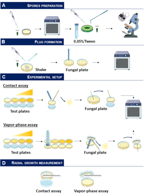
- Prior to the experiment, lay 5 µL of Trichoderma spp. SBT10-2018 spores stored at 4 °C on potato dextrose agar medium (PDA) and incubate for 4 days at 30°C with regular light exposure to promote conidia formation42 (Figure 1, panel A).
NOTE: Trichoderma spp. SBT10-2018 has been isolated from wood and is used as the model in this study for its rapid growth and ease of spore recovery. This strain is preserved by our laboratory. - Recover conidia (Figure 1, panel A)
- Lay 3 mL of 0.05% Tween-20 on the Trichoderma mycelium.
- Use a rake to release conidia from conidiophores; avoid pressing down on the mycelium to prevent hyphae from being torn away.
- Recover the solution rapidly with a micropipette to avoid it being absorbed by the agar medium and transfer into a 15 mL tube.
- Count the total number of spores using a hemocytometer and prepare a solution containing 3 x 10⁶ spores/mL.
NOTE: This step must be performed carefully to prevent hyphae from being extracted. Spore preparation is then checked under microscope. Eventually, for strains presenting highly aerial and fluffy mycelium, a step of filtration using 40 µM strainer filter can be added to eliminate residual mycelium fragment.
2. Fungal plates preparation (Figure 1, panel B)
- Deposit 100 µL of 3 x 10⁶ spores/mL with a micropipette on a 9 cm diameter Petri dish containing PDA medium to obtain 4,800 spores/cm2 corresponding to 925 spores/5 mm diameter-agar plug.
- Add 10 g of 2 mm diameter glass beads with a sterile spatula and perform forward and backward movements parallel and perpendicular to the operator's arm to evenly distribute the spores on the surface of the agar.
- Rotate the plate by 90° and repeat the rotating movements (as in section 2.2); repeat these steps until the plate has been rotated completely.
- Use the plate immediately to set up experiments requiring spores or incubate the plates at 30 °C for 17 h or 24 h when early hyphae or mycelium, respectively, are needed.
NOTE: To compare antifungal activity measured after mycelium plug-transfer and mycelium disk-transfer, use sterile tweezers and place sterile 5 mm cellulose disks randomly onto the surface of the agar plate after spore spreading.
3. Antifungal compounds preparation
- Plant-derived product preparation: garlic-powder preparation
- Peel the cloves of fresh garlic and cut the cloves into 2-3 mm wide slices using a scalpel blade.
- Air-dry the slices for 2 days at 40 °C.
- Grind the slices for 3 x 15 seconds using a knife mill to obtain a fine powder.
- Store the garlic powder at 4 °C in 50 mL tubes before use.
NOTE: As garlic is not autoclaved (to prevent the degradation of temperature-sensitive antifungal compounds) clean the grinder, the scalpel, and the air-dryer with 70% ethanol before use.
- Essential oil preparation
- Prepare 0.5%, 1%, 2.5%, 5% and 20% Thymus vulgaris essential oil solutions in 0.5% Tween-80.
- Mix well to form an emulsion before adding it into the PDA medium (see section 4.2).
- Carbendazim preparation
- Weigh carbendazim to prepare a 200 mg/L ethanol solution (carbendazim is poorly soluble in water).
- Store the solution at room temperature before adding it into the PDA medium (see section 4.2).
CAUTION: Carbendazim presents a health and environmental hazard. Wear gloves and mask when handling this product. Store it in a ventilated space.
4. Contact-inhibition assay
- Preparation of agar plates containing garlic powder
- Prepare and autoclave PDA medium.
- Weigh the desired garlic powder quantity into a 50 mL tube using a sterile spatula, to obtain concentrations generally ranging from 0.25 mg/mL to 16 mg/mL.
- Add 10 mL of PDA after having checked the temperature of the medium on the inside of the wrist. The temperature must be as low as possible to prevent degradation of sensitive molecules. Ideally, this temperature should be 45 °C.
- Homogenize carefully by turning the tube upside down to evenly distribute the powder into the PDA medium. Quickly pour 10 mL into a 5 cm diameter Petri dish (Figure 1, panel C).
- With the Petri dish placed at room temperature, wait until the agar solidifies.
- Preparation of agar plates containing essential oil or carbendazim
- Introduce 10 mL of PDA into a 50 mL tube. Check the temperature as for section 4.1.3.
- Add 100 µL of the different solutions of Thymus vulgaris essential oil in PDA to obtain 0.005%, 0.01%, 0.025%, 0.05% and 0.2% solutions (see section 3.2.1).
- Add the required volume of carbendazim from the 200 mg/L solution to obtain solutions ranging from 0.0625-2 mg/L (see section 3.3.1).
- Homogenize carefully by turning the tube upside down, quickly pour 10 mL into a 5 cm diameter Petri dish (Figure 1, panel C).
- With the Petri dish placed at room temperature, wait until the agar solidifies.
- Contact inhibition assay (Figure 1)
- With a 5 mm diameter sterile stainless-steel tube, plot a circle in the center of Petri dishes containing either PDA or PDA including antifungal compounds. Dispose of the agar cylinder using a sterile toothpick (panel C).
- With a 5 mm diameter sterile stainless-steel tube, plot circles randomly into the fungal plates from section 2. Plot between 15-20 circles per plate (panel B).
- Carefully withdraw the agar-cylinders covered by spores, early hyphae, or mycelium with a sterile toothpick and place the plugs into the empty space of Petri dishes containing either PDA or PDA including antifungal compounds (panel C).
- Incubate the plates containing spores for 48 h at 30 °C, 31 h for the plates containing early hyphae and, 24 h for the plates covered with mycelium (panel C).
- Measure the diameter of radial growth and calculate the percent of fungal-growth inhibition over control using the formula (panel D)
% fungal growth inhibition = (C – A/C)* 100
where C is the diameter of radial growth in PDA medium and A the diameter of radial growth in PDA medium containing the antifungal compounds.
NOTE: To compare antifungal activity measured after mycelium plug-transfer and mycelium disk-transfer, using sterile tweezers, transfer one 5 mm diameter disk previously deposited onto the surface of the fungal plates (section 2 note) at the center of Petri dishes containing either PDA or PDA containing antifungal compounds and proceed exactly as for agar-plug transfer
5. Vapor-Phase inhibition assay
- Preparation of agar plates containing garlic powder
- Proceed as in section 3.1.
- Preparation of agar plate containing essential oil or carbendazim
- Proceed as in section 3.2 and 3.3.
- Preparation of fungal plates
- Proceed as in section 2.
- Vapor-phase antifungal inhibition assay (Figure 1)
- Pour 10 mL of PDA medium into the lid of the 5 cm diameter Petri dishes containing either 10 mL PDA medium or 10 mL of PDA medium containing antifungal compounds into the bottom of the dishes. Wait until complete solidification of the agar at room temperature (panel C).
- Use a 50 mL centrifugal tube as a calibration tool to obtain a circle of PDA in the center of the lid; remove the PDA around the circle with a sterile spatula (panel C).
- Plot a circle in the center of the PDA medium placed into the lid with a 5 mm diameter sterile stainless-steel tube. Discard the agar-cylinder with a sterile toothpick (panel C).
- Form plugs with a 5 mm diameter sterile stainless-steel tube randomly into the fungal plates as in section 4.3.2 (panel B).
- Using a sterile toothpick, carefully transfer the plugs covered either with spores, early hyphae, or mycelium from fungal plates into the lids of assay plates (panel C).
- Incubate at 30 °C as in section 4.3.4 (panel C).
- Measure the diameter of radial growth and calculate the percent of fungal growth inhibition using the formula in section 4.3.5 (panel D).
Representative Results
To evaluate the ability of the quantitative method to discriminate the mode of action of different types of antifungal compounds, we compared the efficacy of three well-known antifungal agents. Carbendazim is a non-volatile synthetic fungicide which has been widely used to control a broad range of fungal diseases in plants39,40. Thymus vulgaris essential oil has been largely described for its antibacterial and antifungal activity and is used as natural food preservative agent41. Garlic powder has been chosen as a model of a plant-derived bioproduct. It has been traditionally used as a natural remedy with antimicrobial activities which have largely been attributed to the presence of volatile organosulfur compounds but also to the presence of non-volatile saponins and phenolic compounds26, giving to this model a complexity relevant in this study.
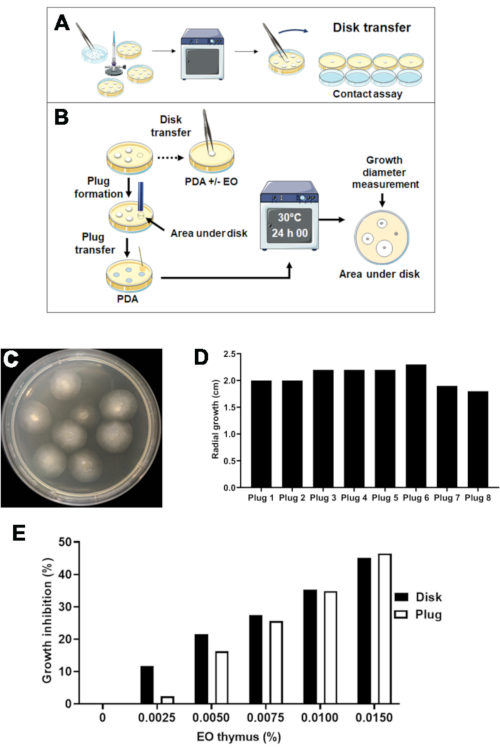
This quantitative method relies on the transfer of agar plugs containing controlled amounts of fungus at different developmental stages from spores to mycelium whereas in the food-poisoned method, 5-day to 7-day old mycelium is transferred from cellulose disks13. In the assay, spores, early hyphae (17 h incubation) and mycelium (24 h incubation)were used as starting fungal material. The use of disk transfer might not be relevant as conidia or residual hyphae remain at least partially on the agar medium after disk transfer, subsequently leading to inaccurate measurement of growth inhibition as illustrated in Figure 2. Different diameters of fungal-radial growth have been observed after transfer of agar areas located under cellulose disks followed by 24 h incubation (Figure 2, panels A, B and C) highlighting the presence of residual fungal hyphae on agar after disk transfer. The quantification of residual hyphae has been confirmed by the measurement of growth leading to up to 22% diameter variability (Figure 2, panel D). The effect on growth inhibition was next evaluated using Thymus vulgaris essential oil as antifungal compound and compared to the inhibition obtained after agar-plug transfer (Figure 2, panel E). Growth inhibition after disk transfer was higher than after agar-plug transfer for low Thymus oil concentrations, leading to an over-estimation of the inhibitory effect, which might be due to incomplete transfer of fungal material and support the approach based on agar-plug transfer.
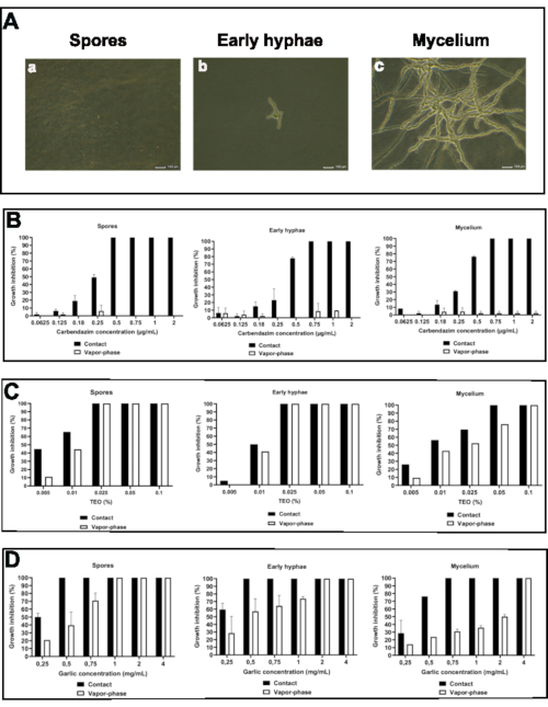
Trichoderma spp. SBT10-2018-growth inhibition triggered by the three antifungal compounds was next evaluated using the contact- and vapor-phase inhibition assays for each fungal stage (Figure 3). Spores were carefully spread on agar plates to obtain 4,800 spores/cm2. They were directly transferred to agar plates containing antifungal compounds through agar-plug extraction using a 5 mm sterile stainless-steel tube, allowing the experiment to start from the spores. For the two other developmental stages, agar plates covered with spores were firstly incubated for 17 h or 24 h at 30°C before transferring the agar plug to allow germination and early development of hyphae (17 h) and mycelium formation (24 h) (Figure 3, panel A). To quantify the contribution of active volatile molecules to the overall antifungal activity, the contact inhibition assay has been adapted and spores, early hyphae, and mycelium were placed at a distance from the antifungal compounds poured into PDA medium as for the contact-inhibition assay. Trichoderma-radial growth was measured over 48 hours and the percentage of inhibition has been determined by comparison to control conditions. The minimum-inhibitory concentration (MIC) has been defined as the lowest concentration of antifungal compounds preventing visible growth after 48 h of incubation at 30°C.
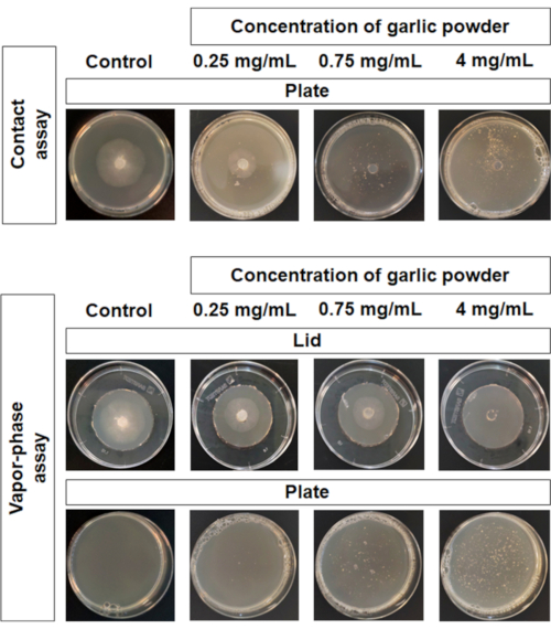
Figure 3(panel B) shows higher spore sensitivity to carbendazim compared to early hyphae and mycelium networks with 50%, 22% and 30% growth inhibition respectively at 0.25 µg/mL carbendazim when Trichoderma and antifungal compounds were in contact. Concomitantly, a MIC value of 0.5 µg/mL has been estimated on spore germination whereas an increase to 0.75 µg/mL has been obtained on early hyphae elongation and mycelium. By contrast, carbendazim had no antifungal effect on Trichoderma when the fungus was placed at distance from the fungicide in accordance with the low volatility of this substance. The results we obtained using Thymus vulgaris essential oil (TEO) as antifungal compound (Figure 3, panel C) have shown a higher spore sensitivity to TEO in comparison to early hyphae and mycelium with 65% and approximately 50% growth inhibition at 0.01% TEO respectively. The MIC values obtained were similar for spore germination and early hyphae elongation (0.025% TEO) and higher for mycelium growth (0.05% TEO). As expected, Thymus vulgaris essential oil presented identical antifungal activity irrespective of the distance beTween the fungus and the oil. Similar MIC values (0.025% TEO) were obtained on spore germination and early hyphae elongation for contact and vapor-phase assays, though at the lower percentage a higher sensitivity has been observed when TEO and Trichoderma spores were in contact (60% growth inhibition in contact versus 45% growth inhibition at distance). Surprisingly, MIC values obtained on the mycelium were different in the contact- and vapor-phase inhibition assays (0.05% versus 0.1%) suggesting that some part of the volatile molecules is not active against a well-developed mycelium. Finally, when using garlic powder as antifungal compound (Figure 3, panel D), a higher efficacy was observed against spore germination (50% growth inhibition at 0.25 mg/mL garlic powder and MIC value of 0.5 mg/mL) and early hyphae elongation (59% growth inhibition at 0.25 mg/mL and MIC value of 0.5 mg/mL) than for mycelium growth (29% growth inhibition at 0.5 mg/mL garlic powder and MIC value of 0.75 mg/mL). When contact- and vapor-phase assays were compared, the results have shown a significant decrease in antifungal activity at distance irrespective of the developmental stage of the fungus. The MIC values moved from 0.5 mg/mL to 1 mg/mL for spore germination, from 0.5 mg/mL to 2 mg/mL for early hyphae elongation, and from 0.75 mg/mL to 4 mg/mL for mycelium growth (Figure 3, panel D and Figure 4 for representative pictures). So, these results suggest that garlic powder contains a mixture of both volatile and non-volatile compounds having antifungal properties.
Altogether, these results show that the relative contribution of volatile and non-volatile agents contained in plant-derived products may be determined at different fungal-growth stages as the experimental conditions are comparable. This approach is then particularly well suited for complex mixtures of antifungal compounds. Thymus vulgaris essential oil is a mixture of volatile compounds and shows a similar activity at distance and in contact for spore germination and early hyphae elongation, supporting the comparison of this vapor-phase and contact-inhibition assay and highlighting that migration into the vapor-phase is not impaired by pouring into the agar medium. The results also underline that garlic powder used as model in this study contains non-volatile active components which have a significant contribution in the overall antifungal activity and which have been neglected in favor of volatile thiosulfinates derived from allium27,28.

Figure 1: Synoptic scheme of the protocol for contact and vapor-phase assays Please click here to view a larger version of this figure.

Figure 2: Inaccuracy associated with fungal transfer from cellulose disk. A. Scheme representing disk transfer onto agar-plates covered by spores and disk transfer onto the surface on agar-plates containing antifungal compounds B. Scheme representing agar transfer of areas under cellulose disks followed by incubation and residual growth measurement. C. Representative picture of radial growth of residual myceliumafter transfer of areas under cellulose disks. D. Radial growth measurement of residual mycelium. E. Effect of Thymus vulgaris essential oil on the growth of mycelium transferred from cellulose disk or agar-plug (N=2, mean ± SD) Please click here to view a larger version of this figure.

Figure 3: Comparison of antifungal activities using the vapor-phase and contact inhibition assays on spores, early hyphae, and mycelium. A. Representative pictures of Trichoderma spores, early hyphae (17 h growth), and mycelium (24 h growth). Trichoderma growth inhibition by carbendazim (B), Thymus vulgaris essential oil (C) and garlic powder (D). (N=2, mean ± SD) Please click here to view a larger version of this figure.

Figure 4: Representative pictures of garlic antifungal activity on agar plates in contact- (A) or vapor-phase (B) inhibition assays Please click here to view a larger version of this figure.
Discussion
The approach presented here allows for the evaluation of antifungal properties of minimally processed plant-derived products. In this protocol, homogenous distribution of spores on the agar surface is achieved using 2 mm glass beads. This step requires handling skills to properly distribute the beads and to obtain reproducible results, ultimately allowing the comparison of antifungal effects at different stages of fungal growth. We found that 5 mm glass beads or excessive rotation while homogenizing during spreading can cause variable growth diameter. Therefore, we recommend training to master spore distribution prior to experimentation. In addition, when plant powders must be tested, attention must be given to homogenous dispersion of the product into the agar medium. To prevent the powder from settling at the bottom of the plate, the product has to be mixed into agar medium when the temperature of the melted medium reaches 45 °C (when room temperature is 24 °C). This temperature has to be adjusted according to the local room temperature to avoid sedimentation.
While the method we describe here can provide valuable insights, a few drawbacks must be considered. This method allows for accurate and side-by-side comparisons in a single experimental setup at the expense of a significant amount of preparation time as the number of agar plates to be prepared can be sizeable depending on the questions that have to be answered. In addition, this assay is a medium-scale assay designed for 5 cm Petri dishes. Therefore, the amount of active substances required to test all the aspects can be substantial. That means that rare substances may not be suitable test candidates for this protocol. A scale-down of the assay can be considered using smaller Petri dishes and reducing the size of the plugs. This could be tested using the benchmarking protocol described here with special attention to the agar-plug extraction, which might be difficult. The accuracy of radial-growth measurements might be reduced at that smaller scale.
Current methods are appropriate for measuring the antifungal activity of compounds in solution and less applicable to studying powders13. The approach we have established is well-adapted for both liquid and solid compounds, which allows evaluation of the antifungal properties of minimally processed plant-derived products. This reduces the time required to test extracts and reduces pitfalls related to active substances displaying poor solubility. As some plant-derived products contain active molecules sensitive to high temperature43, this offers the advantage of limiting the risk of a loss of activity of such compounds. This approach has been adapted from the agar-diffusion method and food-poisoned method15,16,17,18,19 to additionally permit the direct comparison of antifungal activities on different fungal growth stages using similar experimental settings. Agar-plug transfer allows accurate control of the quantities of microorganisms within the assay. This is an advantage over disk transfer, which leads to over-estimation of antifungal effects associated with incomplete transfer of spores or hyphae. Finally, whereas vapor-phase assays are generally not applied to contact inhibition assays27,28,29,30,31, the method we propose addresses the relative contributions of volatile and non-volatile agents contained in complex mixtures such as plant powders or extracts and ultimately allows the evaluation of antifungal activities at different fungal-growth stages.
The approach we describe here might be particularly relevant to support the need for methods that evaluate antifungal properties in plant-derived products for biocontrol. The quantitative method we propose allows operators to determine the respective contributions of volatile and non-volatile compounds to the antifungal activity of a complex bioactive mixture. This provides valuable information that can guide the choice of the modes of application for the treatment and the relevance of performing liquid extraction. Applications that might be considered at a distance from the targeted phytopathogen (for instance, inclusion of the biocontrol product into a packaging) or might need direct contact to optimize the efficacy of the biocontrol product (nebulization onto plants or dipping fruit into a solution of biocontrol product). It also allows the comparison of antifungal efficacy at different fungal-growth stages from spore germination to later stage mycelium growth, which leads to the definition of recommendations for establishing control thresholds required to apply biocontrol products to crops. Indeed, defining the efficacy of the substances at different fungal stages may help to categorize whether substances can be used as preventive or curative treatments and to plan the schedule for plant-treatments with biocontrol products. This is essential to leverage the efficacy of the products when used in field or post-harvest.
Açıklamalar
The authors have nothing to disclose.
Acknowledgements
We are very grateful to Frank Yates for his precious advice. This work was supported by Sup'Biotech.
Materials
| Autoclave-vacuclav 24B+ | Melag | ||
| Carbendazim | Sigma | 378674-100G | |
| Distilled water | |||
| Eppendorf tubes | Sarstedt | 72.706 | 1.5 mL |
| Falcons tubes | Sarstedt | 547254 | 50 mL |
| Five millimeters diameter stainless steel tube | retail store | / | |
| Food dehydrator | Sancusto | six trays | |
| Garlic powder | Organic shop | ||
| Glass beads | CLOUP | 65020 |  2 mm 2 mm |
| Hemocytometer counting cell | Jeulin | 713442 | / |
| Incubator | Memmert | UM400 | 30 °C |
| Knife mill | Bosch | TSM6A013B | |
| Manual cell counter | Labbox | HCNT-001-001 | / |
| Measuring ruler | retail store | ||
| Microbiological safety cabinets | FASTER | FASTER BHA36, TYPE II, Cat 2 | |
| Micropipette | Mettler-Toledo | 17014407 | 100 – 1000 µL |
| Micropipette | Mettler-Toledo | 17014411 | 20 – 200 µL |
| Micropipette | Mettler-Toledo | 17014412 | 2 – 20 µL |
| Petri dish | Sarstedt | 82-1194500 |  55 mm 55 mm |
| Petri dish | Sarstedt | 82-1473 |  90 mm 90 mm |
| Pipette Controllers-EASY 60 | Labbox | EASY-P60-001 | / |
| Potato Dextrose Agar | Sigma | 70139-500G | |
| Precision scale-RADWAG | Grosseron | B126698 | AS220.R2-ML 220g/0.1mg |
| Rake | Sarstedt | 86-1569001 | / |
| Reverse microscope AE31E trinocular | Grosseron | M097917 | / |
| Sterile graduated pipette | Sarstedt | 1254001 | 10 mL |
| Thymus essential oil | Drugstore | Essential oil 100% | |
| Tips 1000 µL | Sarstedt | 70.762010 | |
| Tips 20 µL | Sarstedt | 70.760012 | |
| Tips 200 µL | Sarstedt | 70.760002 | |
| Tooth pick | retail store | ||
| Trichoderma spp strain | Strain of LRPIA laboratory | ||
| Tween-20 | Sigma | P1379-250ML | |
| Tween-80 | Sigma | P1754-1L | |
| Tweezers | Labbox | FORS-001-002 | / |
Referanslar
- FAO. Global food losses and food waste – Extent, causes and prevention. FAO. , (2011).
- da Cruz Cabral, L., Fernández Pinto, V., Patriarca, A. Application of plant compounds to control fungal spoilage and mycotoxin production in foods. International Journal of Food Microbiology. 166 (1), 1-14 (2013).
- Romanazzi, G., Smilanick, J. L., Feliziani, E., Droby, S. Postharvest biology and technology integrated management of postharvest gray mold on fruit crops. Postharvest Biology and Technology. 113, (2016).
- Morton, V., Staub, T. A Short History of Fungicides. APSnet Feature Articles. (1755), 1-12 (2008).
- Brandhorst, T. T., Klein, B. S. Uncertainty surrounding the mechanism and safety of the post- harvest fungicide Fludioxonil. Food and Chemical Toxicology. 123, 561-565 (2019).
- Bénit, P., et al. Evolutionarily conserved susceptibility of the mitochondrial respiratory chain to SDHI pesticides and its consequence on the impact of SDHIs on human cultured cells. PLoS ONE. 14 (11), 1-20 (2019).
- Usall, J., Torres, R., Teixidó, N. Biological control of postharvest diseases on fruit: a suitable alternative. Current Opinion in Food Science. 11, 51-55 (2016).
- Tripathi, P., Dubey, N. K. Exploitation of natural products as an alternative strategy to control postharvest fungal rotting of fruit and vegetables. Postharvest Biology and Technology. 32 (3), 235-245 (2004).
- Abbey, J. A., et al. Biofungicides as alternative to synthetic fungicide control of grey mould (Botrytis cinerea)-prospects and challenges. Biocontrol Science and Technology. 29 (3), 241-262 (2019).
- Soylu, E. M., Kurt, &. #. 3. 5. 0. ;., Soylu, S. In vitro and in vivo antifungal activities of the essential oils of various plants against tomato grey mould disease agent Botrytis cinerea. International Journal of Food Microbiology. 143 (3), 183-189 (2010).
- Liu, S., Shao, X., Wei, Y., Li, Y., Xu, F., Wang, H. Solidago canadensis L. essential oil vapor effectively inhibits botrytis cinerea growth and preserves postharvest quality of strawberry as a food model system. Frontiers in Microbiology. 7, 0 (2016).
- El-Mogy, M. M., Alsanius, B. W. Cassia oil for controlling plant and human pathogens on fresh strawberries. Food Control. 28 (1), 157-162 (2012).
- Balouiri, M., Sadiki, M., Ibnsouda, S. K. Methods for in vitro evaluating antimicrobial activity: A review. Journal of Pharmaceutical Analysis. 6 (2), 71-79 (2016).
- Arikan, S. Current status of antifungal susceptibility testing methods. Medical Mycology. 45 (7), 569-587 (2007).
- Girmay, Z., Gorems, W., Birhanu, G., Zewdie, S. Growth and yield performance of Pleurotus ostreatus (Jacq. Fr.) Kumm (oyster mushroom) on different substrates. AMB Express. 6 (1), 87 (2016).
- Fischer, M. S., Glass, N. L. Communicate and fuse: how filamentous fungi establish and maintain an interconnected mycelial network. Frontiers in Microbiology. 10, 1-20 (2019).
- Mohana, D. C., Raveesha, K. A. Anti-fungal evaluation of some plant extracts against some plant pathogenic field and storage fungi. Journal of Agricultural Technology. 4 (1), 119-137 (2007).
- Balamurugan, S. In vitro activity of aurantifolia plant extracts against phytopathogenic fungi phaseolina. International Letters of Natural Sciences. 13, 70-74 (2014).
- Ameziane, N., et al. Antifungal activity of Moroccan plants against citrus fruit pathogens. Agronomy for sustainable development. 27 (3), 273-277 (2007).
- Rizi, K., Murdan, S., Danquah, C. A., Faull, J., Bhakta, S. Development of a rapid, reliable and quantitative method – “SPOTi” for testing antifungal efficacy. Journal of Microbiological Methods. 117, 36-40 (2015).
- Imhof, A., Balajee, S. A., Marr, K., Marr, K. New methods to assess susceptibilities of Aspergillus isolates to caspofungin. Mikrobiyoloji. 41 (12), 5683-5688 (2003).
- Goussous, S. J., Abu el-Samen, F. M., Tahhan, R. A. Antifungal activity of several medicinal plants extracts against the early blight pathogen (Alternaria solani). Archives of Phytopathology and Plant Protection. 43 (17), 1745-1757 (2010).
- Ng, T. B. Antifungal proteins and peptides of leguminous and non-leguminous origins. Peptides. 25 (7), 1215-1222 (2004).
- Hu, Z., Zhang, H., Shi, K. Plant peptides in plant defense responses. Plant Signaling and Behavior. 13 (8), (2018).
- Iriti, M., Faoro, F. Chemical diversity and defence metabolism: How plants cope with pathogens and ozone pollution. International Journal of Molecular Sciences. 10 (8), 3371-3399 (2009).
- Lanzotti, V., Bonanomi, G., Scala, F. What makes Allium species effective against pathogenic microbes. Phytochemistry Reviews. 12 (4), 751-772 (2013).
- Kyung, K. H. Antimicrobial properties of allium species. Current Opinion in Biotechnology. 23 (2), 142-147 (2012).
- Hyldgaard, M., Mygind, T., Meyer, R. L. Essential oils in food preservation: mode of action, synergies, and interactions with food matrix components. Frontiers in microbiology. 3, 12 (2012).
- Bueno, J. Models of evaluation of antimicrobial activity of essential oils in vapour phase: a promising use in healthcare decontamination. Natural Volatiles & Essential Oils. 2 (2), 16-29 (2015).
- Doi, N. M., Sae-Eaw, A., Suppakul, P., Chompreeda, P. Assessment of synergistic effects on antimicrobial activity in vapour- and liquidphase of cinnamon and oregano essential oils against Staphylococcus aureus. International Food Research Journal. 26 (2), 459-467 (2019).
- Amat, S., Baines, D., Alexander, T. W. A vapour phase assay for evaluating the antimicrobial activities of essential oils against bovine respiratory bacterial pathogens. Letters in Applied Microbiology. 65 (6), 489-495 (2017).
- Feyaerts, A. F., et al. Essential oils and their components are a class of antifungals with potent vapour-phase-mediated anti-Candida activity. Scientific Reports. 8 (1), 1-10 (2018).
- Wang, T. H., Hsia, S. M., Wu, C. H., Ko, S. Y., Chen, M. Y., Shih, Y. H., Shieh, T. M., Chuang, L. C. Evaluation of the antibacterial potential of liquid and vapor phase phenolic essential oil compounds against oral microorganisms. PLoS ONE. 11 (9), 1-17 (2016).
- Dean, R., et al. The Top 10 fungal pathogens in molecular plant pathology. Molecular Plant Pathology. 13 (4), 414-430 (2012).
- Leadbeater, A. Recent developments and challenges in chemical disease control. Plant Protection Science. 51 (4), 163-169 (2015).
- Jin, C., Zeng, Z., Fu, Z., Jin, Y. Oral imazalil exposure induces gut microbiota dysbiosis and colonic inflammation in mice. Chemosphere. 160, 349-358 (2016).
- Kumar, R., Ghatak, A., Balodi, R., Bhagat, A. P. Decay mechanism of postharvest pathogens and their management using non-chemical and biological approaches. Journal of Postharvest Technology. 6 (1), 1-11 (2018).
- Talibi, I., Boubaker, H., Boudyach, E. H., Ait Ben Aoumar, A. Alternative methods for the control of postharvest citrus diseases. Journal of Applied Microbiology. 117 (1), 1-17 (2014).
- Arya, R., Sharma, R., Malhotra, M., Kumar, V., Sharma, A. K. Biodegradation Aspects of Carbendazim and Sulfosulfuron: Trends, Scope and Relevance. Current Medicinal Chemistry. 22 (9), 1147-1155 (2015).
- European Food Safety Authority. Conclusion on the peer review of the pesticide risk assessment of the active substance carbendazim. EFSA Journal. 8 (5), 1-76 (2010).
- Sakkas, H., Papadopoulou, C. Antimicrobial activity of basil, oregano, and thyme essential oils. Journal of Microbiology and Biotechnology. 27 (3), 429-438 (2017).
- Steyaert, J. M., Weld, R. J., Mendoza-Mendoza, A., Stewart, A. Reproduction without sex: conidiation in the filamentous fungus Trichoderma. Mikrobiyoloji. 156, 2887-2900 (2010).
- Leontiev, R., Hohaus, N., Jacob, C., Gruhlke, M. C. H., Slusarenko, A. J. A Comparison of the antibacterial and antifungal activities of thiosulfinate analogues of allicin. Scientific Reports. 8 (1), 1-19 (2018).
- Scorzoni, L., et al. The use of standard methodology for determination of antifungal activity of natural products against medical yeasts Candida sp and Cryptococcus sp. Brazilian Journal of Microbiology. 38 (3), 391-397 (2007).

