Deacetylation Assays to Unravel the Interplay between Sirtuins (SIRT2) and Specific Protein-substrates
Özet
This protocol describes the required steps to execute in vitro and in vivo deacetylation assays in order to establish the role of proteins as specific deacetylation substrates for sirtuins and further study the role of reversible – lysine acetylation as a post-translational modification.
Abstract
Acetylation has emerged as an important post-translational modification (PTM) regulating a plethora of cellular processes and functions. This is further supported by recent findings in high-resolution mass spectrometry based proteomics showing that many new proteins and sites within these proteins can be acetylated. However the identity of the enzymes regulating these proteins and sites is often unknown. Among these enzymes, sirtuins, which belong to the class III histone lysine deacetylases, have attracted great interest as enzymes regulating the acetylome under different physiological or pathophysiological conditions. Here we describe methods to link SIRT2, the cytoplasmic sirtuin, with its substrates including both in vitro and in vivo deacetylation assays. These assays can be applied in studies focused on other members of the sirtuin family to unravel the specific role of sirtuins and are necessary in order to establish the regulatory interplay of specific deacetylases with their substrates as a first step to better understand the role of protein acetylation. Furthermore, such assays can be used to distinguish functional acetylation sites on a protein from what may be non-regulatory acetylated lysines, as well as to examine the interplay between a deacetylase and its substrate in a physiological context.
Introduction
Post-translational modifications (PTMs) regulate cell signaling networks allowing cells to rapidly respond to internal and external signals. Over the last few decades, many different PTMs playing a pivotal role in diverse processes have been identified but only a few have been studied extensively, such as phosphorylation, acetylation and ubiquitination 1-3. Focusing on acetylation, Allfrey et al. were the first to propose a role for histone acetylation in regulating gene transcription about 50 years ago 4. Research in this field has revealed that histone lysine acetylation modulates chromatin condensation and it is considered to be an epigenetic mark as part of the histone code 5. Although it took a long time until the discovery of tubulin as the first non-histone acetylation target 6, it is well established now that hundreds of eukaryotic proteins beyond histones can be acetylated and lysine acetylation has been recognized as a wide-spread PTM that may rival phosphorylation and ubiquitination in its prevalence 7-9. Interestingly, non-histone acetylated proteins can be signaling molecules in the cytoplasm, transcription factors in the nucleus, and metabolic enzymes in mitochondria, highlighting the significance of acetylation in regulating a plethora of cellular processes.
The acetylation status of a protein depends on the coordinated and opposing function of lysine acetyltransferases (KATs) and lysine deacetylases (KDACs) which add and remove acetyl groups from proteins. The reversible acetylation of lysine, which involves neutralization of a positive charge 10, alters protein structure and it seems very likely to also alter enzymatic function in several cases 11-13. Focusing on KDACs, 18 proteins have been identified in the human and mouse genomes 14-16. Among them, mammalian sirtuins (also called class III histone lysine deacetylases) which are distinct from other members as they require NAD+ for their enzymatic function, have attracted extensive interest in this research field 16. In mammals, seven sirtuins (SIRT1-7) have been identified, each of them sharing a conserved 275-amino-acid catalytic core domain, which are mainly categorized according to their subcellular localization to the nucleus (SIRT1, 6, and 7), mitochondria (SIRT3, 4, and 5), or cytoplasm (SIRT2). SIRT1-3 have a robust deacetylation activity, while SIRT4 is reported to display ADP-ribosyltransferase activity, SIRT5 may function as a protein desuccinylase and demalonylase, and SIRT6 and SIRT7 display weak deacetylase activity but are involved in other types of acylations 17. In accordance with the significance of acetylation as a regulatory PTM modification involved in several cellular functions, sirtuins have also been implicated in a wide range of processes. After the first breakthrough studies establishing the role of sirtuins in life span extension, it has been shown that they are involved in diverse cellular functions including DNA repair, maintenance of genomic instability, apoptosis, response to stress and inflammation, control of energy efficiency, circadian clocks and metabolism, as well as contributing to the initiation and/or progression of age-related diseases such as cancer, neurodegeneration and type 2 diabetes 15,16.
Despite the significant progress in the field of sirtuin biology, more work remains to unravel undiscovered roles and functions through the identification of novel substrates. This is evidenced more emphatically by recent advances in high-resolution mass spectrometry (MS) based proteomics which have significantly increased the number of proteins found to be acetylated but most importantly have identified several different acetylated lysines in each protein, arguing that acetylation may be as wide-spread as other PTMs such as phosphorylation 7,8,17. Taking into consideration that specific deacetylases have not yet been identified for most of these acetylated proteins-substrates, it is reasonable to suggest that both in vitro and in vivo deacetylation assays are needed to confirm and establish an acetylated protein as a legitimate substrate of a specific deacetylase. In the experimental protocols described below, details will be given on how to perform both in vitro and in vivo deacetylation assays using SIRT2 as the specific deacetylase.
Protocol
1. In Vitro Deacetylation Assay
- Purification of SIRT2
- Prepare a 10 cm culture dish of HEK 293T cells cultured in 8 ml DMEM with 10% FBS and antibiotics and grown in a 37 °C, 5% CO2 tissue culture incubator to be about 70% confluent the next day.
- The next day, co-transfect 4 µg pCDH-puro-GFP-SIRT2-Flag (lentivector made in our lab), 4 µg pCMV- dR8.2 dvpr (packaging vector) and 0.5 µg pCMV-VSV-G (envelope vector) 18 using polyethylenimine (PEI) at a ratio of 3 µl PEI/µg DNA.
- Change media after 12 hr.
- Collect supernatant after 36-48 hr.
- Remove debris by centrifugation at 1,000 x g for 5 min at 4 °C.
- After filtering through 0.22 µm filters, make 1 ml aliquots of SIRT2 lentivirus and keep at -80 °C.
- Thaw SIRT2 lentivirus on ice (it is important to store the virus at -80 °C when the virus is not used).
- Infect a 6-well plate of 80-90% confluent HEK 293T cells with 1 ml of lentivirus in the presence of 8 µg/ml polybrene.
- Place virus-infected cells in 37 °C incubator until the next day (24 hr).
- Replace medium after 24 hr.
- 48 hr after infection, check infection efficiency by detecting GFP positive cells under a fluorescent microscope.
- If infection efficiency is good (at least 40-50% infected cells), replace medium with culture medium containing 2 µg/ml puromycin to select for stable SIRT2 over expressing cells.
Note: This takes approximately 2 weeks, and the medium is changed every 3-4 days. Non-infected cells die over this selection period and only infected cells with the SIRT2 lentivirus grow. - Analyze expression of Flag-tagged SIRT2 by western blotting using an anti-Flag antibody (1:1,000 dilution) after running 40 µg of total protein on a 10% SDS-PAGE gel before starting the purification process (recommended) 19,20.
- To purify active SIRT2, split cells in 1:3 ratio by trypsinization in order to have enough dishes of HEK 293T cells stably overexpressing Flag-SIRT2 (Ten 10 cm dishes will allow purification of enough protein for subsequent experiments).
- To trypsinize the cells, remove culture medium and eliminate residual serum by rinsing cells with sterile DPBS. Slowly add 2.5 ml of a 0.25% Trypsin-EDTA solution to cover the cell monolayer, incubate at room temperature for 30-45 sec. After removing the Trypsin-EDTA solution, incubate at 37 °C until cells start to detach. Then add culture medium and transfer cells to new dishes.
- Remove culture medium, wash cells twice in PBS and collect cells by trypsinization followed by centrifugation at 1,000 x g for 10 min at 4 °C.
- Lyse cell pellet in 500 µl lysis buffer A (20 mM HEPES pH 7.9, 18 mM KCl, 0.2 mM EGTA, 1.5 mM MgCl2, 20% Glycerol, 0.1% NP-40) including a protease inhibitors cocktail and 1 µM TSA for 30 min at 4 °C under constant rotation.
- Centrifuge at 14,000 x g for 15 min at 4 °C and transfer supernatant to a new tube.
- Perform immunoprecipitation using an anti-Flag antibody conjugated to agarose beads as described below.
- Add 200-300 µl of anti-Flag antibody conjugated to agarose beads to the protein lysate (10-20 mg total protein).
- Incubate overnight at 4 °C with constant agitation.
- Collect immunoprecipitates by centrifugation at 1,000 x g for 5 min at 4 °C.
- Wash the pellet 5 times with 10 ml lysis buffer A at 1,000 x g for 5 min at 4 °C. After each wash, pellet the beads and replace the supernatant with buffer A.
- Prepare 1x Flag peptide solution (including a protease inhibitors cocktail and 1 µM TSA in PBS).
- Add 500 µl of 1x Flag peptide solution to the agarose beads and incubate for 30 min at 4 °C under constant rotation.
- Collect supernatant by centrifugation at 6,000 x g for 2 min at 4 °C.
- Repeat previous two steps.
- Remove beads from supernatant by using filter tubes and centrifugation at 15,000 x g for 1 min at 4 °C.
- Concentrate eluted sample until the final protein concentration is 1 µg/µl using an ultrafiltration membrane.
- Check purification process by performing electrophoresis using 1 µg of the eluted sample.
- Stain gel with a commercially available staining solution to confirm SIRT2 purification.
- Keep purified SIRT2 at -80 °C.
- Purification of Acetylated Protein-substrate
- Prepare 10 cm x 10 cm culture dishes with HEK 293T cells to be about 70% confluent the next day.
- The next day, co-transfect cells with 5 µg of a Flag tagged plasmid vector expressing the protein-substrate as well as 5 µg of the specific acetyl-transferase which can acetylate the protein using PEI at a ratio of 3 µl PEI/µg DNA.
Note: If the specific acetyl-transferase is not known, co-transfect with a mixture of known histone acetyl-transferases (HATs) such as p300, CBP, GCN5, Tip60 and PCAF. - Change medium after 12 hr.
- The day before lysing the cells, treat the cells with 1 µM TSA and 2 mM nicotinamide (NAM) overnight by replacing the culture medium with medium containing TSA/NAM to maximize acetylation levels of the protein-substrate by inhibiting deacetylases.
- 48 hr after transfection, remove culture medium, wash cells twice in PBS and collect cells by trypsinization followed by centrifugation at 1,000 x g for 10 min at 4 °C.
- Lyse cell pellet in 500 µl lysis buffer A per dish (20 mM HEPES pH 7.9, 18 mM KCl, 0.2 mM EGTA, 1.5 mM MgCl2, 20% Glycerol, 0.1% NP-40) including a protease inhibitors cocktail, 1 µM TSA and 2 mM NAM for 30 min at 4 °C under constant rotation.
- Centrifuge at 14,000 x g for 15 min at 4 °C and transfer supernatant to a new tube.
- Perform immunoprecipitation using an anti-Flag antibody conjugated to agarose beads as described below.
- Add 200-300 µl of anti-Flag antibody conjugated to agarose beads to the protein lysate (10-20 mg total protein).
- Incubate overnight at 4 °C with constant agitation.
- Collect immunoprecipitates by centrifugation at 1,000 x g for 5 min at 4 °C.
- Wash the pellet 2 times with 10 ml lysis buffer A at 1,000 x g for 5 min at 4 °C. After each wash, pellet the beads and replace the supernatant with lysis buffer A.
- Wash the pellet 3 times with 10 ml lysis buffer A without NAM at 1,000 x g for 5 min at 4 °C. After each wash, pellet the beads and replace the supernatant with lysis buffer A. It is important not to carry over any NAM to the deacetylation reaction.
- Prepare 1x Flag peptide solution (including protease inhibitors and 1 µM TSA in PBS).
- Add 500 µl of 1x Flag peptide solution to the agarose beads and incubate for 30 min at 4 °C under constant rotation.
- Collect supernatant by centrifugation at 6,000 x g for 2 min at 4 °C.
- Repeat previous two steps.
- Remove beads from supernatant by using filter tubes and centrifugation at 15,000 x g for 1 min at 4 °C.
- Using an ultrafiltration membrane, concentrate eluted sample until the protein concentration is 1 µg/ul.
- Perform electrophoresis by running 1 µl of the eluted sample on a 4-12% SDS-PAGE gel.
- Confirm acetylation of protein-substrate by western blotting using antibodies against Ac-K (1:1,000 dilution) and the protein-substrate (dilution depends on the specific antibody used).
- Keep purified acetylated protein-substrate at -80 °C.
- In Vitro Deacetylation Reaction
- Prepare the deacetylation buffer B (50 mM Tris-HCl, pH 7.5, 150 mM NaCl, 1 mM MgCl2 0.5 µM TSA)
- Prepare 3 different reactions in different tubes in 20 µl final volume (Table 1).
- Include additional reactions to confirm deacetylation of the protein-substrate by SIRT2 (optional). Add 2 µg of purified deacetylation null mutant SIRT2 (this can be done using the protocol described in 1.1 after using a pCDH-puro-GFP-SIRT2H187Y-Flag lentivector) to the reaction instead of deacetylase active SIRT2 or add 10 mM nicotinamide (NAM) to the reaction to inhibit the deacetylation activity of SIRT2 (Table 1).
- Incubate reactions for 3 hr at 30 °C under constant agitation.
- Stop the reaction by adding 20 µl of 2x sample buffer and boil the samples for 10 min at 95 °C.
- Perform electrophoresis by running the reaction mixture on a 4-12% SDS-PAGE gel and detect the acetylation status of the protein-substrate by western blotting using an anti Ac-K antibody (1:1,000 dilution) 19,20.
2. In Vivo Deacetylation Assay
- SIRT2 Overexpression in Cultured Cells
- Prepare 10 cm culture dishes with HEK 293T cells stably overexpressing pCDH-puro-GFP-empty vector, pCDH-puro-GFP-SIRT2-Flag and pCDH-puro-GFP-SIRT2H187Y-Flag (as described in 1.1) to be about 70% confluent.
- Alternatively, prepare 10 cm culture dishes with cells about 70% confluent and perform transient overexpression using 5 µg of the vectors mentioned above using PEI at a ratio of 3 µl PEI/µg DNA.
- Perform western blotting of total cellular extracts using an anti-Flag antibody (1:1,000 dilution) to demonstrate overexpression of Flag-tagged SIRT219.
- RNA Interference to Knockdown SIRT2 in Cultured Cells
- Prepare a 10 cm culture dish of HEK 293T cells to be about 70% confluent the next day.
- The next day, co-transfect 4 µg pLKO1-puro-sh SIRT2 (lentivector) 19, 4 µg pCMV- dR8.2 dvpr (packaging vector) and 0.5 µg pCMV-VSV-G (envelope vector) 18 using polyethylenimine (PEI) at a ratio of 3 µl PEI/µg DNA.
Note: For knocking down SIRT2, we use simple hairpin shRNAs in the pLKO.1 lentiviral vector designed by The RNAi Consortium (TRC). - Follow the procedure as described in 1.1 to produce shSIRT2 lentivirus and infect target cells to knockdown SIRT2.
- Perform western blotting of total cellular extracts using an anti-SIRT2 antibody (1:1,000 dilution) to demonstrate efficient SIRT2 knockdown after running 40 µg of total protein on a 10% SDS-PAGE gel.
- Alternatively, prepare 10 cm culture dishes with cells about 70% confluent and perform transient knockdown of SIRT2 using siRNAs following manufacturer's instructions.
- Perform western blotting of total cellular extracts using an anti-SIRT2 antibody (1:1,000 dilution) to demonstrate efficient SIRT2 knockdown after running 40 µg of total protein on a 10% SDS-PAGE gel.
- Immunoprecipitation and Immunoblotting to Detect Acetylated Levels in Cultured Cells
- 12 hr before collecting cell pellets from either cells overexpressing SIRT2 (see 2.1 above) or SIRT2 knocked down cells (see 2.2 above), add 1 µm TSA to the culture medium to inhibit non Class III histone deacetylases.
- Remove culture medium, wash cells twice in PBS and collect cells by trypsinization followed by centrifugation at 1,000 x g for 10 min at 4 °C.
- Add 10 ml lysis buffer A (20 mM HEPES pH 7.9, 18 mM KCl, 0.2 mM EGTA, 1.5 mM MgCl2, 20% Glycerol, 0.1% NP-40) including a protease inhibitors cocktail, 1 µM TSA and 2 mM NAM.
- Incubate at 4 °C for 30 min under constant rotation.
- Collect supernatants by centrifugation at 20,000 x g for 15 minutes at 4 °C.
- Calculate protein concentration by using a Bradford protein assay.
- Transfer 1 mg of total protein to new tubes in a final volume of 1 ml using lysis buffer A. Use Table 2 as a reference to prepare protein samples from cells either overexpressing SIRT2 or after SIRT2 knockdown.
- Perform an immunoprecipitation using an antibody against the specific protein-substrate together with Protein A/G (40 µl bead slurry is recommended). Alternatively, use an anti Ac-K antibody conjugated to agarose beads (20 µl bead slurry is usually enough) to immunoprecipitate all acetylated proteins.
- Incubate samples at 4 °C under constant rotation overnight.
- Collect immunoprecipitates by centrifugation at 1,000 x g for 2 min at 4 °C.
- Wash beads 5 times with 1 ml buffer A at 1,000 x g for 2 min at 4 °C.
- Resuspend beads in 40 µl of 2x sample buffer.
- Boil samples for 10 min at 95 °C.
- Perform electrophoresis by running the eluted samples (40 µl from step 2.3.12) on a 4-12% SDS-PAGE gel without disturbing the beads while transferring the eluted samples.
- Detect acetylated levels of the protein-substrate by Western blotting using an anti-Ac-K antibody (1:1,000 dilution) if a protein-substrate specific antibody was used for immunoprecipitation, or a specific antibody against the protein-substrate (follow recommendations for the specific antibody regarding dilution) if anti Ac-K beads were used for the immunoprecipitation.
Representative Results
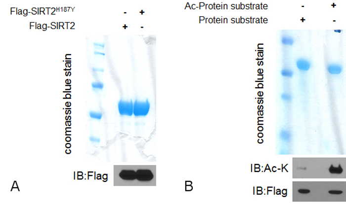
In order for a protein to be considered as a legitimate deacetylation target for any enzyme with deacetylation activity, both in vitro and in vivo deacetylation assays need to be performed to establish the interplay between the deacetylase and its substrate. For the in vitro deacetylase assay, the purification of both the deacetylase and the acetylated protein substrate is required before the assay can be done. Here we use the cytoplasmic sirtuin SIRT2 as the specific deacetylase to be studied. SIRT2 is a deacetylase enzyme and the use of a mutant which lacks the deacetylase activity is highly recommended as a negative control for the deacetylation assay. Using the purification procedure described in 1.1, both SIRT2 and SIRT2H187Y can be successfully purified at a final concentration of 1 µg/µl. This can be confirmed after running the purified proteins on a gel followed by staining (Figure 1A, upper) as well as after western blotting using an anti-Flag antibody since both SIRT2 and SIRT2H187Y are tagged with a Flag peptide (Figure 2A, lower). The same purification procedure can be followed for the purification of the protein substrate with some modifications. The protein needs to be acetylated before it can be used for the deacetylation assay which means that it needs to be purified in cells overexpressing the specific acetyl transferase or a mixture of HATs able to acetylate the protein. Using the purification procedure described in 1.2, the acetylated protein can be purified (Figure 1B, upper). To confirm that the protein is indeed acetylated and can be used for the in vitro deacetylation assay, western blotting using an anti Ac-K antibody is necessary to show increased acetylated levels of the purified protein in cells overexpressing the acetyl-transferases (Figure 1B, lower).
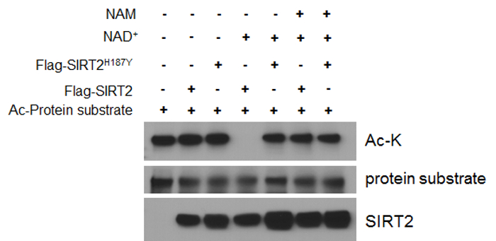
After successful protein purification, the in vitro deacetylation assay can be performed. Sirtuins, including SIRT2, are class III histone lysine deacetylases which are distinct from other deacetylases as they require NAD+ for their enzymatic function. Consistent with this, no deacetylation can be detected by western blotting using an anti Ac-K antibody in the absence of NAD+, regardless of the presence of catalytically active SIRT2 (Figure 2, lane 2 vs 1). On the contrary, decreased acetylated levels of the acetylated protein when both SIRT2 and NAD+ are present in the reaction suggest that the protein can be considered as a deacetylation target for SIRT2. In order to verify these results, different negative controls can be used. For example no difference in the deacetylation levels of the protein can be detected when a deacetylation deficient SIRT2 (SIRT2H187Y) is used instead of the catalytically active wild-type SIRT2 (Figure 2, lane 5 vs 4). Similar effect can be seen when a well-established sirtuin inhibitor, NAM, is added to the reaction mixture, implying that the decrease in the acetylated levels of the protein is mediated by the deacetylase activity of SIRT2.
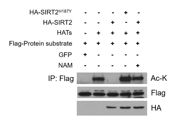
Aberrant deacetylation is involved in several cellular processes and age-related diseases. Thus, a crucial step in deepening our knowledge regarding the interplay between a deacetylase and its substrate is to establish this interconnection in cells where this phenomenon may have a physiological impact. Moreover, checking deacetylation in cell culture systems in vivo allows the study of the deacetylation event in a cell or tissue specific context which can be more easily connected to a specific phenotype or outcome. This excludes the possibility that the detected in vitro deacetylation activity is artificial due to the presence of both the deacetylase and its substrate in the tube at the same time, which may never happen under normal physiological conditions in cells. For the in vivo deacetylation assay, cells overexpressing both SIRT2 and SIRT2H187Y as well as the protein substrate can be used following the procedures described in 2.1 and 2.2. Cell extracts prepared from these cells can be confirmed to express SIRT2, SIRT2H187Y, and the protein substrate by western blotting using specific antibodies. In the assay described here, all exogenously expressed proteins are tagged for ease of the performed experiments (Figure 3, lower panel to detect HA-tagged SIRT2/SIRT2H187Y and middle panel to detect Flag-tagged protein substrate). To determine whether the target protein is a deacetylation substrate of SIRT2, immunoprecipitation is performed using an anti-Flag antibody to pull down the target protein followed by western blotting using an anti Ac-K antibody. Decrease in the acetylated levels of the protein in cells expressing SIRT2 (Figure 3 upper panel, lane 3 vs 2) and failure to affect acetylated levels in cells expressing the deacetylase deficient SIRT2 mutant (Figure 3 upper panel, lane 4 vs 3) suggest that the acetylated protein can be a deacetylation target of the specific deacetylase in vivo. This can be confirmed further when SIRT2-mediated deacetylation is inhibited after treating cells with NAM (Figure 3 upper panel, lane 5 vs 3) establishing the specific deacetylase-substrate axis in the studied cells.

Figure 1: Purification of both the deacetylase and the acetylated protein-substrate to be used for the in vitro deacetylation assay. (A) 1 µg of both SIRT2 and SIRT2H187Y (deacetylation defective mutant) were run on a gel following the purification protocol as described in 1.1. The gel was stained with a commercially available staining solution and after destaining, both purified proteins can be detected (upper). Proteins can be detected after transferring in PVDF membrane and western blotting using an anti-Flag antibody (lower). (B) 1 µg of protein substrate and hyper acetylated protein substrate (alpha-enolase was used as a SIRT2 substrate based on mass spectrometry unpublished data generated in our lab) were run on a gel following the purification protocol as described in 1.2. The gel was stained with a commercially available staining solution and after destaining, the purified protein substrate can be detected (upper). Acetylation can be detected after transferring in PVDF membrane and western blotting using an anti-Ac-K antibody (middle). Total proteins can be detected using an anti-Flag antibody (lower). Please click here to view a larger version of this figure.

Figure 2: In vitro deacetylation assay. Purified acetylated protein substrate (alpha-enolase) is incubated with SIRT2 or SIRT2H187Y (deacetylation defective mutant) in the presence of NAD+ (lanes 4 and 5). Decreased acetylated levels are detected by western blotting using an anti Ac-K antibody in the presence of SIRT2 but not in the presence of SIRT2H187Y (lane 4 vs 5). No deacetylation activity is observed when NAD+ is not included in the reaction mixture (lane 2 vs 4) or when NAM is added to the reaction mixture (lane 6 vs 4). Please click here to view a larger version of this figure.

Figure 3: In vivo deacetylation assay. HEK293T cells stably overexpressing either SIRT2 or SIRT2H187Y were co-transfected with the protein substrate (alpha-enolase) and HATs to increase the acetylated levels of the protein (lane 2 vs 1). Deacetylation was checked in vivo after immunoprecipitation using an anti-Flag antibody to pull down the protein substrate and western blotting using an anti Ac-K antibody. Acetylation is significantly decreased in the SIRT2 overexpressing cells as compared to the SIRT2H187Y overexpressing cells (lane 3 vs 4). The SIRT2-mediated deacetylation of the protein substrate is inhibited when cells are treated with NAM (lane 5 vs 3). Please click here to view a larger version of this figure.
| Reactions | 1 | 2 | 3 | 4 (optional) | 5 (optional) |
| purified acetylated protein-substrate (10 μg) | + | + | + | + | + |
| buffer B | + | + | + | + | + |
| purified SIRT2 (2 μg) | – | + | + | – | + |
| NAD+ (1 mΜ) | – | – | + | + | + |
| purified SIRT2H187Y (2 μg) | – | – | – | + | – |
| NAM (10 mM) | – | – | – | – | + |
Table 1: Reaction ingredients.
| samples | overexpression | knockdown |
| 1 | pCDH-puro-GFP-empty vector | pLKO1 empty vector or si ctr |
| 2 | pCDH-puro-GFP-SIRT2-Flag | pLKO1 sh SIRT2 1 or si SIRT2 1 |
| 3 | pCDH-puro-GFP-SIRT2H187Y-Flag | pLKO1 sh SIRT2 2 or si SIRT2 2 |
Table 2: Protein samples.
Discussion
Recent high throughput proteomic studies have established acetylation as a widespread PTM found not only in nucleus but also in cytoplasm and mitochondria 7,8,21-23. Taking into account the likelihood that many more acetylated proteins and sites might have been not detected due to several reasons, such as specificity of the anti-Ac-K antibodies used, the low abundance of the acetylated proteins, and the transient nature of the PTM, it is safe to predict that more acetylated proteins remain to be discovered in the future, highlighting the importance of reversible acetylation as a regulatory modification directing diverse cellular processes. Regardless of the magnitude of acetylation, identifying the deacetylases which can specifically deacetylate such protein-substrates is an important first step in unraveling the biology of protein acetylation and understanding the complexity of this phenomenon. Towards this direction, use of both in vitro and in vivo deacetylation assays are crucial and necessary to establish the regulatory interplay between deacetylases and their substrates.
Here we present an example of how deacetylation assays can be used in order for an acetylated protein to be considered as a legitimate deacetylation target of SIRT2. As it has already been mentioned, the sirtuin family of proteins consists of seven members which can be found in the cytoplasm, mitochondria, and nucleus 15. Given the increasing interest regarding the role of these proteins in the different subcellular compartments which can be associated with distinct cellular processes, the above described deacetylation assays can be used with slight modifications to identify new substrates for all different members of the sirtuin family. For example, when mitochondrial proteins are used in deacetylation assays related to SIRT3, it may be necessary to include a mitochondrial isolation step 24,25 between cell lysis and immunoprecipitation during the purification process for the in vitro deacetylation assay or during the in vivo deacetylation assay before detecting acetylated protein levels. In a similar way, when nuclear proteins need to be used as deacetylation targets in these assays, nuclear extracts following subcellular fractionation might be used for protein purification and the in vivo deacetylation assay.
A critical step for executing the in vitro deacetylation assay is the successful purification of both the deacetylase and the acetylated protein-substrate. Verification of the cells overexpressing the deacetylase as well as the acetylated protein-substrate before the initiation of the purification step is highly recommended. Furthermore, detection of the purified proteins after running a small amount of the isolated proteins on an SDS-PAGE gel before the in vitro deacetylation reaction is also recommended. This will not only confirm the completion of the purification step but will help investigators to focus their efforts on optimizing the deacetylation reaction conditions for any given protein-substrate and deacetylase of interest in case of an unsuccessful deacetylation assay which may require changes in the enzyme-substrate ratio used. Of note, even if a positive deacetylation reaction is detected using the in vitro assay, further validation is required before any conclusion can be made regarding the establishment of a protein as a deacetylation target of a specific deacetylase. This limitation is due to the fact that the co-presence of the substrate and the deacetylase in the tube does not mean that both can be found in the same complex in cells or tissues. This implies that the deacetylation needs to be further validated in vivo. With the focus on the in vivo deacetylation assay, it is worth mentioning that the steady state acetylation levels of a given protein under the experimental conditions tested is a crucial factor in determining whether overexpression or knockdown experiments are more suitable to establish the functional interplay between a deacetylase and its substrate. High levels of acetylation may direct the experimental design towards overexpressing the specific deacetylase to detect a decrease in the acetylation levels. On the contrary, when low levels of acetylation are detected under normal conditions, increased acetylation after knocking down the specific deacetylase might establish the mechanistic connection between a deacetylase and its substrate. Taking all these together, it is clear that the deacetylation assays described in this protocol are necessary to link a deacetylase with its potential substrate.
The next challenges in the field of lysine acetylation dictate that deacetylation assays will be used in the future to address some of the missing links. The considerable gap between the large number of identified mitochondrial lysine sites and the few with a validated regulatory function 26 highlights the need to distinguish bona fide functional acetylation sites from what may be widespread spurious modifications. In vitro and in vivo deacetylation assays carried over by specific deacetylases may be further combined with small scale MS analysis to reveal specific target lysines on a tested substrate to distinguish functional acetylation sites from what may be non-regulatory acetylated lysines. In this regard, sites which are deacetylated due to the enzymatic activity of a sirtuin are more likely to play a functional role compared to sites that are not deacetylated. Another significant challenge is the examination of lysine acetylation in a physiological context. It seems very likely that the acetylation profile of a cell may vary under specific experimental conditions. In vivo deacetylation assays in cells under specific experimental conditions can provide further insight into the connection between the deacetylase and the potential substrate in physiological and pathophysiological scenarios depending on the cell type, or the exposure to specific signaling molecules and stressors. Such studies can establish the functional role of a deacetylation event in a physiological context and connect it to a phenotypic outcome or response. More importantly, deacetylation assays in combination with small scale MS analysis can further provide meaningful information regarding regulatory acetylated lysines under specific cellular conditions.
In conclusion, identification of the enzymes that deacetylate acetylated proteins and sites by using both in vitro and in vivo deacetylation assays will help to resolve the complexity of the acetylome and will contribute to better understanding of the regulatory role of acetylation under various physiological conditions.
Açıklamalar
The authors have nothing to disclose.
Acknowledgements
The project described here was supported by a grant from NIH/NCI (NCI-R01CA182506-01A1) as well as by the Robert H. Lurie Comprehensive Cancer Center – The Lefkofsky Family Foundation/Liz and Eric Lefkofsky Innovation Research Award to A.V. We would like to thank members of the laboratory (Carol O'Callaghan and Elizabeth Anne Wayne) for critical reading and editing this manuscript.
Materials
| cell culture dishes | Denville Scientific Inc. | T1110 and T1115 | |
| pCDH-puro-GFP lentiviral vector | System Biosciences | CD513B-1 | |
| pCMV- dR8.2 dvpr (packaging vector) | Addgene | 8455 | |
| pCMV-VSV-G (envelope vector) | Addgene | 8454 | |
| polyethylenimine (PEI) | Polysciences Inc. | 24885 | other transfection reagents can be used as well. PEI is cost effective and very efficient in transfecting 293T cells |
| 0.22μm filters | Denville Scientific Inc. | F5512 | |
| polybrene | Sigma | H9268 | |
| fluorescent microscope | Carl Zeiss MicroImaging Inc. | Axiovert 200 | |
| puromycin | Invivogen | A11138-03 | |
| PBS | Corning | 21-031-CM | |
| anti-Flag antibody | Sigma | F3165 | |
| HEPES | Sigma | H3375 | |
| KCl | Sigma | P9541 | |
| Glycerol | Sigma | G5516 | |
| NP-40 | Sigma | 74385 | |
| MgCl2 | Sigma | M9272 | |
| EGTA | Sigma | 34596 | |
| protease inhibitors coctail 100x | Biotool | B14001 | |
| Trichostatin A (TSA) | Sigma | T8552 | selective inhibitor of class I and II histone deacetylases (HDACs) but not class II HDACs (sirtuins) |
| anti-Flag agarose beads | Sigma | A2220 | |
| centrifuge | Eppendorf | 5417R | |
| rotator | Thermo Scientific | 415220Q | |
| filter tubes | Millipore | UFC30HV00 | |
| Flag peptide | Sigma | F3290 | |
| Vivaspin Centrifugal Concentrator | Sartorius Stedim Biotech S.A. | VS0102 | |
| SimplyBlue SafeStain solution | Invitrogen | LC6060 | |
| NuPAGE LDS sample buffer (4x) | Life Techologies | NP0007 | |
| anti-Ac-K antibody | Cell Signaling | 9441 | several anti-Ac-K antibodies are available. In our hands, the Cell Signaling antibody exhibits the highest sensitivity |
| Tris-HCl pH 7.5 | Sigma | T5941 | |
| NAD+ | Sigma | N0632 | required cofactor for sirtuins |
| nicotinamide (NAM) | Sigma | 72340 | selective inhibitor class II HDACs (sirtuins) |
| pLKO.1 lentiviral vector | Addgene | 8453 | |
| SIRT2 si RNA | Qiagen | GS22933 | |
| anti-SIRT2 antibody | Proteintech | 15345-1-AP | |
| Bradford protein assay | BIO-RAD | 500-0006 | |
| anti Ac-K agarose beads | Immunechem | ICP0388 |
Referanslar
- Hunter, T. The age of crosstalk: phosphorylation, ubiquitination, and beyond. Molecular Cell. 28, 730-738 (2007).
- Gao, M., Karin, M. Regulating the regulators: control of protein ubiquitination and ubiquitin-like modifications by extracellular stimuli. Molecular Cell. 19, 581-593 (2005).
- Philp, A., Rowland, T., Perez-Schindler, J., Schenk, S. Understanding the acetylome: translating targeted proteomics into meaningful physiology. American Journal of Physiology. Cell Physiology. 307, C763-C773 (2014).
- Allfrey, V. G., Faulkner, R., Mirsky, A. E. Acetylation and Methylation of Histones and Their Possible Role in the Regulation of Rna Synthesis. Proceedings of the National Academy of Sciences of the United States of America. 51, 786-794 (1964).
- Shahbazian, M. D., Grunstein, M. Functions of site-specific histone acetylation and deacetylation. Annual Review of Biochemistry. 76, 75-100 (2007).
- L’Hernault, S. W., Rosenbaum, J. L. Chlamydomonas alpha-tubulin is posttranslationally modified in the flagella during flagellar assembly. The Journal of Cell Biology. 97, 258-263 (1983).
- Kim, S. C., et al. Substrate and functional diversity of lysine acetylation revealed by a proteomics survey. Molecular Cell. 23, 607-618 (2006).
- Choudhary, C., et al. Lysine acetylation targets protein complexes and co-regulates major cellular functions. Science. 325, 834-840 (2009).
- Smith, K. T., Workman, J. L. Introducing the acetylome. Nature Biotechnology. 27, 917-919 (2009).
- Roth, S. Y., Denu, J. M., Allis, C. D. Histone acetyltransferases. Annual Review of Biochemistry. 70, 81-120 (2001).
- Ozden, O., et al. Acetylation of MnSOD directs enzymatic activity responding to cellular nutrient status or oxidative stress. Aging. 3, 102-107 (2011).
- Anderson, K. A., Hirschey, M. D. Mitochondrial protein acetylation regulates metabolism. Essays in Biochemistry. 52, 23-35 (2012).
- Schwer, B., Bunkenborg, J., Verdin, R. O., Andersen, J. S., Verdin, E. Reversible lysine acetylation controls the activity of the mitochondrial enzyme acetyl-CoA synthetase 2. Proceedings of the National Academy of Sciences of the United States of America. 103, 10224-10229 (2006).
- Haberland, M., Montgomery, R. L., Olson, E. N. The many roles of histone deacetylases in development and physiology: implications for disease and therapy. Nature Reviews. Genetics. 10, 32-42 (2009).
- Finkel, T., Deng, C. X., Mostoslavsky, R. Recent progress in the biology and physiology of sirtuins. Nature. 460, 587-591 (2009).
- Houtkooper, R. H., Pirinen, E., Auwerx, J. Sirtuins as regulators of metabolism and healthspan. Nature reviews. Molecular Cell Biology. 13, 225-238 (2012).
- Choudhary, C., Weinert, B. T., Nishida, Y., Verdin, E., Mann, M. The growing landscape of lysine acetylation links metabolism and cell signalling. Nature Reviews. Molecular Cell Biology. 15, 536-550 (2014).
- Stewart, S. A., et al. Lentivirus-delivered stable gene silencing by RNAi in primary cells. RNA. 9, 493-501 (2003).
- Kim, H. S., et al. SIRT2 maintains genome integrity and suppresses tumorigenesis through regulating APC/C activity. Cancer Cell. 20, 487-499 (2011).
- Zhang, H., et al. SIRT2 directs the replication stress response through CDK9 deacetylation. Proceedings of the National Academy of Sciences of the United States of America. 110, 13546-13551 (2013).
- Weinert, B. T., et al. Proteome-wide mapping of the Drosophila acetylome demonstrates a high degree of conservation of lysine acetylation. Science Signaling. 4, ra48 (2011).
- Still, A. J., et al. Quantification of mitochondrial acetylation dynamics highlights prominent sites of metabolic regulation. The Journal of Biological Chemistry. 288, 26209-26219 (2013).
- Sadoul, K., Wang, J., Diagouraga, B., Khochbin, S. The tale of protein lysine acetylation in the cytoplasm. Journal of Biomedicine & Biotechnology. 2011, 970382 (2011).
- Preble, J. M., et al. Rapid isolation and purification of mitochondria for transplantation by tissue dissociation and differential filtration. Journal of Visualized Experiments : JoVE. , e51682 (2014).
- Garcia-Cazarin, M. L., Snider, N. N., Andrade, F. H. Mitochondrial isolation from skeletal muscle. Journal of Visualized Experiments : JoVE. , (2011).
- He, W., Newman, J. C., Wang, M. Z., Ho, L., Verdin, E. Mitochondrial sirtuins: regulators of protein acylation and metabolism. Trends in Endocrinology and Metabolism: TEM. 23, 467-476 (2012).

