Structural Analyses of An Epidermal Growth Factor Receptor-Specific Single-Chain Fragment Variable via An In Silico Approach
概要
This antibody homology modeling prediction protocol is followed by antibody-receptor Pyrx docking and molecular dynamic simulation. These three primary methods are used to visualize the accurate antibody-receptor binding areas and the binding stability of the final structure.
Abstract
Single-chain fragment variable (scFv) antibodies were previously constructed of variable light and heavy chains joined by a (Gly4-Ser) 3 linker. The linker was created using molecular modeling software as a loop structure. Here, we introduce a protocol forin silico analysis of a complete scFv antibody that interacts with the epidermal growth factor receptor (EGFR). The homology modeling, with Pyrx of protein-protein docking and molecular dynamic simulation of the interacting scFv antibody and EGFR First, the authors used a protein structure modeling program and Python for homology modeling, and the antibody scFv structure was modeled for homology. The investigators downloaded Pyrx software as a platform in the docking study. The Molecular dynamic simulation was run using modeling software. Results show that when the MD simulation was subjected to energy minimization, the protein model had the lowest binding energy (-5.4 kcal/M). In addition, the MD simulation in this study showed that the docked EGFR-scFv antibody was stable for 20-75 ns when the movement of the structure increased sharply to 7.2 Å. In conclusion, in silicoanalysiswas performed, and the molecular docking and molecular dynamics simulations of the scFv antibody proved the effectiveness of the designed immune-therapeutic drug scFv as a specific drug therapy for EGFR.
Introduction
Conformational changes in the protein (ligand and receptor) always occur based on structure-based functions. The study of the possible binding grooves of the protein and prediction of the stable binding interaction is an advanced method to prepare drugs for better use in the human body. Homology modeling followed by docking and molecular dynamic simulation is a straightforward method for accurate prediction of stable interactions of binding between the residues of receptors and constructed antibodies that are used as specific personalized medicine1,2. The predicted model structure can show conformational changes and rearrangements in ligand-receptor binding sites, particularly at the antibody-receptor interface. There are many reasons for these changes, such as the rotation of side chains, global structural transformation, or more complex modifications. The main reason for homology modeling is to distinguish a protein’s tertiary structure from its primary structure2,3.
A tyrosine kinase receptor called epidermal growth factor receptor (EGFR) plays many biological roles in cancer cells, including apoptosis4,5, differentiation6,7, cell cycle progression8,9, development9,10, and transcription11. EGFR is one of the well-known therapeutic targets for breast cancer12. The overexpression of regular kinase activity such as EGFR usually leads to cancer cell progression, which can be repressed by many kinds of cancer inhibitors13. The epidermal growth factor receptor (EGFR) was used as a receptor for the single chain fragment variable specifically constructed to work against this receptor. Its predicted structure was used to test the antibody binding activity.
In this paper, the scFv antibody structure was modeled using modeling software with Python script and the homology modeling method14,15. A homology model can be built from the protein and amino acid sequences of receptors and ligands16,17. Additionally, advanced bioinformatics technologies such as molecular docking were employed to predict how small molecule ligands will bind to the correct target binding site. The docking would balance the development of novel drugs directed toward multiple diseases. The binding behavior is taken into consideration5,18.
Furthermore, molecular docking is a critical technique to facilitate and speed up ligand-receptor binding development. Molecular docking enables scientists to virtually screen a library of ligands against a target protein and predict the binding conformations and affinities of the ligands to the target receptor protein. Molecular dynamic simulation (MNS) demonstrates how the residues move in space, simulates the antibody motions toward their receptors, and finally informs antibody design efforts. This study is a novel prediction of grid box dimensions that decided how the scFv antibody binds to EGFR and the detection of the energy and time of that binding in MDsimulation.
Protocol
1. Secondary structure predictions of a single chain fragment variable (scFv) protein
- Build the single-chain fragment variable (scFv) protein's 3D structure with BLAST protein data bank (PDB), KABAT numbering, and the modeling software. The scFv consists of a linker (Gly4-Ser) that connects a variable heavy chain (VH) and a variable light chain (VL).
- Use the molecular modeling software to build the linker as a loop structure, and perform all these methods as described in previous studies2,19,20.
2. Template selection and scFv and EGFR 3D structure prediction and homology modeling
- Choose template 1ivo for EGFR structures (based on its high resolution). Download the 1ivo.pdb file from the pdb website, as shown in Figure 1B.
- Prepare the input 1ivo.pdb file as described below.
- In the 1ivo.pdb file, remove all external ligands by opening the pdb.org website and selecting the 1ivo. Structure, and looking for the name of the ligands under the small molecule title on the 1ivo structure page of the pdb website.
- Find the ligand name NAG. Open the 1ivo.pdb file downloaded from the pdb website and find the termination residue (TER.).
- Delete the residues of the external ligands in the 1ivo structure, starting from the residue after TER. and before the residue ends. Save the 1ivo.pdb file on the system.
- Prepare the saved 1ivo.pdb file as described below.
- Download the Autodock docking software (autodock.scripps.edu) from the window selection area. Click on the Open 1ivo.pdb File.
- Use the Edit command to choose Add Hydrogen > Add, then select Polar Only, and then press Ok.
- Use the Edit command to add Kollman charges (Supplementary Figure 1). Use the Edit command to delete water. Save the 1ivo.pdb file on the pc.
- Minimize the energy of the 1ivo.pdb structure as described below.
- Download SPDBV. software from http://spdbv.vital-it.ch/disclaim.html. Open the 1ivo.pdb file.
- Select all. Select the command Perf > Energy minimization > Ok (Supplementary Figure 2). Save the 1ivo file on the pc.
- Prepare the full model scFv using homology modeling as described below.
- Download the modeling software17 and the Python script 3.7.9 shell from the Window- 64. Keep the downloaded software files in the D drive.
- Prepare the input files as described below.
- Load the scFv Pdb file in fasta format from the NCBI website and rename the file TARGET.ali. as described in Supplementary Coding File 1. Choose the template using the Blast section in NCBI, paste the sequenced file, select in pdb format 7det.pdb as described in Supplementary Coding File 2, and then submit. Then, use the pdb.org website to obtain the template file.
- Prepare the third input file as align2d.py (Python) as described in Supplementary Coding File 3, which opens as shown in Supplementary Figure 3A. Press the Show More Option, then go to Edit with IDLE > Edit with EDLE (64-bit). Run using the run module 5 command in the align2d.py to obtain two output files: Tar- 7det.ali and Tar- 7det.pap.
- Complete the previous three steps to use the command in the last input file.
- Add a new input file model-single.py (command python file) as shown in Supplementary Coding File 4 and as described below.
- Press the Show More Option, then go to Edit with IDLE > Edit with EDLE3.7 (64-bit). Run using the (run module 5) command as shown in Supplementary Figure 3B.
NOTE: The resulting output files are the six files of the homology models shown in Supplementary Figure 3C.
- Press the Show More Option, then go to Edit with IDLE > Edit with EDLE3.7 (64-bit). Run using the (run module 5) command as shown in Supplementary Figure 3B.
3. Receptor secondary structure prediction and evaluation
- Detect the homology models' correction and accuracy as described below.
- Create the Ramachandran plot for the scFv models and EGFR models by downloading the visualization tool from https://discover.3ds.com/discovery-studio-visualizer-download.
- Open the file, then right-click with the mouse and select the display sequence (Supplementary Figure 4). Copy the sequence and paste it into the Pictorial database of 3D structures (pdbsum) www.ebi.ac.uk/thornton-srv/databases/pdbsum/.
- Select search by sequence, paste the copy of the sequence, and then submit it. Create the plot as shown in Figure 1B,D.
4. Protein-protein docking
- Download the virtual screening tool software.
- Go to File> Read Molecules > Load 1ivo.pdb. Right-click on the protein in the autodock panel to make a macromolecule. Right-click again to make a ligand (Supplementary Figure 5).
- Click in the autodock panel to select protein and then select ligand.
- Open the protein list. Then, from the list, select scFv Protein.
- Go to Toggle Selection Spheres. Adjust the grid box to the center of the receptor. Click Forward when the round pink button appears.
- To prepare the pdbqt files for both scFv-antibody and EGFR (1ivo) structures, use the following steps.
- Go to C Drive > Program Files (86) > Users, then choose the pyrx file that contains macromolecules and protein output files that were saved as a pdbqt file.
- Then, save the single-chain fragment variable (scFv) antibody pdbqt file.
- Download PyMOL software at PyMOL | pymol.org. Use PyMOL software to show the scFv antibody-receptor EGFR configurations.
- Go to file and open C:Usersilham.mgltoolsPyRxMacromoleculesprotein. Prepare the docking configurations of the scFvantibodyinteracting with the receptor in Figure 2A as described below.
- Use the display option to show the (1ivo)-receptor file as sequence residues with a white background shown in Supplementary Figure 6.
- Display the docking configuration file with higher resolution to see the ligand color in green and red residue colors. Display the (1ivo)-receptor rigid surface in yellow.
- Go to file and open C:Usersilham.mgltoolsPyRxMacromoleculesprotein. Prepare the docking configurations of the scFvantibodyinteracting with the receptor in Figure 2A as described below.
- Prepare the docking configurations of the scFvantibodyinteracting with the receptor in Figure 2B, as described below.
- Download the docking software from the window selection area. Use Autodock to show the scFv antibody-receptor EGFR configurations and conformations.
- In Autodock, choose the Analyze Option, then open the Autodock Vina result. Go to File and open C:Usersilham.mgltoolsPyRxMacromoleculesprotein.
- Select the protein receptor pdb file, then select the area of the ligand configuration (scFv antibody structure). Connect the rigid surface of the receptor with the configuration ofthe docked structure and hide the rest of the receptor. Hide the far residues of the receptor from the connected residues to the ligand, as shown in Figure 2B.
- The protein-protein complex was then considered ready to perform MD simulation.
- Download the docking software from the window selection area. Use Autodock to show the scFv antibody-receptor EGFR configurations and conformations.
5. Molecular dynamic simulation (MD simulation) of the EGFR-scFv antibody docking complex
- Download the MD simulation software and use it as follows.
- Prepare the EGFR (1ivo) pdb file using the reparation wizard as in Supplementary Figure 7A. Operate the preprocess section to refine the file. Send the refined file to be set in the system builder.
- Load the molecular dynamics simulation software from the working directory. Add the ions and upgrade the refined file to reach 20 Å to submit the job (Table 1), also shown in Supplementary Figure 7B.
- Load the EGFR (1ivo) pdb from the imported file, then choose 100 ns timesteps to run it (Supplementary Figure 7C).
- Start the analysis of the simulation after the completion of the MDsimulation as described below.
- Create a job folder and save it in the cms file . Load the cms file to perform this step in the MD simulation.
- Create a working directory for project folders and report the energy values. Use S.I.D. pdf to report the simulation, as shown in Figure 3A, and the interaction diagram and H bond, as shown in Figure 3B.
- Load the pdf file of cms by browsing from the folder and use TIP3P as the model for file volume minimization.
- Create the solvation file to perform this step shown in Supplementary Figure 7D. Save pdf file through the software, and analyze the data, resulting in Figure 4, Figure 5, and Figure 6.
- Generate an MDsimulation finalization setup by creating the resolve file. Find the results in the boundary box, as shown in Figure 7.
Representative Results
Using phage display technology, the scFv gene anti-EGFR was created from the mouse B-cell hybridoma line C3A820,21. The single chain fragment variable (scFv) structure models of the VH and VL structures were built separately, according to Chua et al.22. After that, the models were visible as ribbons produced using RasMol. Then, using molecular modeling software, a synthetic peptide [Gly4Ser)3 was used to join the separately modeled VH and VL structures. The ligand interaction with the receptor is triggered as a local structure that changes the binding sites called variable ends. The complementarity-determining region (CDR) found in antibody protein structures) creates a natural grooving binding for epitope binding3,23. Antigenic binding causes a CDR conformational change to fit residue 183 with the ideal shape and facilitate the proper physicochemical properties of the residues. Instead of comparing the crystal structures of the antibody-bound and antibody-unbound molecules, a dynamic examination of the binding process is thought to be the best way to understand the underlying allosteric 3D mechanism23.
As shown in Figure 1A-D, the plot is used to determine whether the conformation of the backbone is correctly predicted. As shown in Figure 1A,C, most of the peptides are in the correct center of the plot, demonstrating the accuracy and correctness of the structures in Figures 1B,D. The models are subsequently evaluated using Ramachandran plots to determine the accuracy of these models24. The full scFv model-building and evaluation process were prepared so that the average energy profile was below zero. The two Ramachandran plots showed the correct backbone confirmation for the receptor template of 3njp and scFv antibody template of 1osq. The majority of peptides are centralized β sheets24, while the rest are in the α helix of the plot25. The Ramachandran plot is the fastest and easiest method to detect structural peptide correction and accuracy24,26.
Figure 2A shows the crystal structure of the binding interaction between the receptor and ligand in the surface structure. Figure 2B shows the interaction profile of confirmation and lowest binding. Protein-protein docking was performed with PyRx software, and the ligand was kept flexible while the protein structure was kept rigid. The macromolecule and the ligand were prepared with the aid of the MGL 1.5.7 tools program. The ligand was synthesized by rotation to make it flexible, and the macromolecule was created by removing water molecules and adding hydrogen atoms. Antibody-bound and antibody-unbound 186 crystal structures were used to store the potential energy arising from the interaction between each atom in the flexible ligand13. Within the functional domain of the chosen protein molecule, the ligand was docked around the discovered conserved region16. The preferred interaction was selected based on the favorable binding conformation between the macromolecule and ligand that had the lowest binding energy (B.E.), as determined by the docking program25,26. The grid box size was 40 Å x 40 Å x 40 Å along the X, Y, and Z axes and centered at 90.653, 56.2181, and 50.1986, whereas the targeted docking grid box size was set to 40 Å x 40 Å x 40 Å and centered at the X, Y, and Z coordinates of 49.086, 25.0, and 25.0, respectively. The size of the grid box was generated manually.
Figure 3A shows the ligand-receptor residue interaction (receptor interaction). As shown, the dark green residues show the residues that had stronger binding between the scFv antibody-EGFR (1ivo) with H-bonds. In contrast with the red residues with different types of bonding, as shown in Figure3A, the total number of residues is 1117, defined as A=513, B=510, C=7, and D =447. A is the receptor, there are 8685 heavy atoms, and the interaction charges are +55. Furthermore, eight hydrogen bond interactions are shown in Figure 3B: 1H-bond interaction of DNA. Protein with asn:32 of protein receptor, 1 H-bond interaction of DNA. protein with asn:33 of protein receptor, 4 H-bonds with 3tpo, 1 H-bond with tp 3p45, and 1H-bond with t3p5627. Therefore, these interactions exhibit very good docked complex stability and strength.
As shown in Figure 3B, the hydrogen bonds for docking stability (ligand-receptor interaction) and the number of detected hydrogen bonds always determine the strength of docking stability. Protein folding is made more likely by hydrogen bonds, and the chain has access to all degrees of freedom to achieve its ideal structure. However, for stiff binding, the protein molecules are already folded27,28,29 , leaving the chains with just six degrees of freedom in translation and rotation to arrange themselves into the most advantageous bound configuration.
Molecular dynamics simulations (MDS) of EGF receptor binding with scFv protein were observed. In Table 1, counter ion/salt information, applying the NaCl method to EGF receptor binding with the scFv protein binding interaction, and root-mean-square deviation (RMSD) can be used to detect regions of metastable states in addition to the stable state. The authors observed that the predicted structures are not stable, and there is a possibility that the trajectory obtained by a simulation is not reliable. Therefore, the MD simulation can show the consistent movement pattern of the trajectory obtained from the docking of the receptor-antibody interaction, resulting in a steering force that produces stable values in the predicted structures.
As shown in Table 1, the computations are carried out with NaCl concentrations ranging from C = 0.5 m to C = 0.75 m. Analyses are performed on how ions affect the hydrogen258-linked water network and the tetrahedral structure30. Because of a non-tetra-dihedral configuration, the analysis demonstrates that increasing salt content lowers the tetrahedral arrangement of water31. We identify the origin and angular properties of this nontetrahedral structure using the nearest neighbor method. Furthermore, the tetrahedral structure of water is more significantly impacted by the comparison ions Na+ and Cl– than by other ions30,31. The RMSD approach is the standard method. In the RMSD, the protein is located on the Y-axis. The protein frames and the structural backbone's reference frame frequently line up. When hydrogen bonds are estimated in close proximity to the most stable structure of the Trp-cage, the RMSD is calculated32,33.
As shown in Figure 4, the root mean square deviation (RMSD) was calculated; the protein RMSD increased gradually from 3.2 Å to 6 Å for the first 0 to 20 ns time step. Then, from 20 to 75 ns, the protein RMSD was maintained in the range 4 Å to 5.6 Å and then changed from 75 ns to 90 ns. Finally, the protein RMSD movements increased sharply until 7.2 Å and decreased until 6.4 Å up to the last timestep. This indicates the free movement of the proteins. This shows the instability of the docked structure when the time step increases, and the stability of the receptor-ligand docked structure is provided when the time step of RMSD is in the range of 20 to 75 ns. The above plot shows that the RMSD evaluates the structural conformation throughout the simulation. RMSD analysis indicated that the simulation was balanced and stable.
Root mean square fluctuation (RMSF), is shown in Figure 5. In the RMSF, a particular atom's displacement was compared to a reference structure, and the average number of atoms was measured. It is frequently used to discern whether a structure is stable in the time scale of the simulations or not. Furthermore, in Figure 5, the protein shows an RMSF in the range of 2.4 Å for residues between 1-100. Then, it decreases to 2a for 200-300 residues; furthermore, it fluctuates to 5.6 Å or 500 residues and then sharply declines to 2.4 Å for residues until 1000 ns. Finally, there is one sharp peak at the 1000 position with a decrease until 2.4 Å at the terminal. The RMSF results help characterize changes in the ligand atom positions during docking25. These results represent the binding process, and usually, the final model, the closest model, represents the crystal structure in terms of RMF, and binding interactions can be shown clearly in final images, as demonstrated in the previous study by Huang et al.2.
As observed in34, the docked structure of the EGFR-scFv antibody was stable over 1000 ns of MDsimulation. Therefore, protein secondary structure elements (SSEs), such as alpha helices and beta-strands, were monitored throughout the simulation to observe the changes in the residue index in the formation of protein SSEs35,36. Assignment over time is shown in Figure 6. Protein secondary structure elements (SSEs), such as α-helices (Figure 6 in red) and β-strands (Figure 6 in blue), are monitored throughout the simulation. The plot above reports the SSE distribution by residue index throughout the protein structure. As shown in Figure 7, the cartoon model shows protein segments, lipids, and water molecules arranged in straight lines and the lipids' phosphorus atoms in beads.
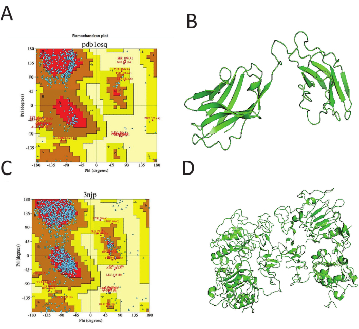
Figure 1: Plot for template selection. The plot is used to determine whether the backbone conformation is correctly predicted. As shown in (A) and (B), most of the peptides are in the correct center of the plot, demonstrating the accuracy and correctness of the structures in (C) and (D). Please click here to view a larger version of this figure.
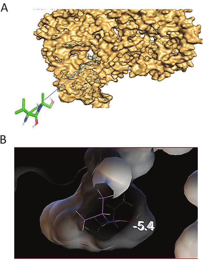
Figure 2: Docking structure and configuration. (A) The Docking structure of the receptor-scFv antibody shows the crystal structure of binding interaction between receptor and ligand in surface structure. (B) Docking configuration. Docking configuration Interaction profile of confirmation and lowest binding presented. Please click here to view a larger version of this figure.
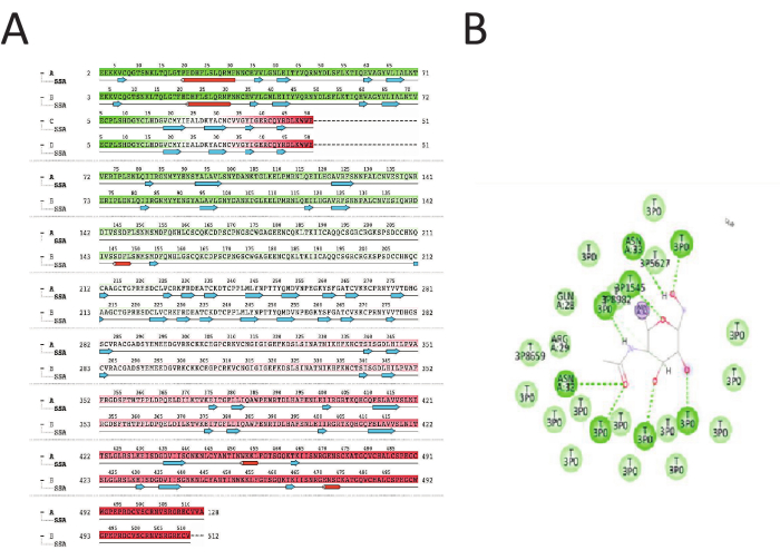
Figure 3: Ligand-receptor residues interaction. (A) Residues interaction of Ligand-receptor structure shows the total number of residue numbers is 1117 residues defined to A =513, B=510, C=7, D =447. A is the receptor. The number of heavy atoms is 8685, while the interaction charges are +55. The dark green alignment is the strongest interaction with the H-bonds. Ligand-receptor residues alignment shows that the green is the strongest bond with the H-bond, the light green is a lighter bond, and the red is the weaker bond. (B) The ligand atoms interact with the protein residues. The Interactions between the ligand and receptor protein occur in more than 30.0% of the simulation time. The receptor residues are connected with the H bond of the ligand atoms. The receptor residues in dark green circles represent the strongest H-bond interaction between the EGFR receptor and scFv antibody. There are eight H bonds, which are considered a strong interaction. Please click here to view a larger version of this figure.
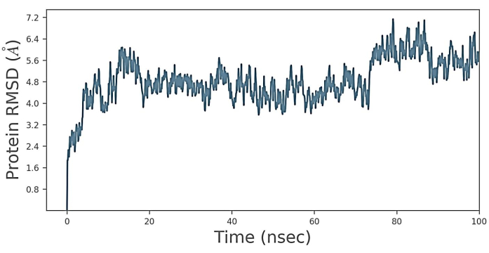
Figure 4: Root mean square deviation (RMSD). RMSD plot of receptor-ligand complex for the 20 ns simulation time. It is observed that protein forms 3.2 Å to 6 Å for the first 0 to 20 ns time step. Additionally, the protein, from 20 ns to 75 ns time step moved. The movement is maintained from 4 Å to 5.6 Å and 75 to 90 ns. The movement increases sharply to 7.2 Å and then decreases to 6.4 A until the last timestep. These results proved that the docked structure EGFR-scFv antibody was stable over the 1000 ns of MD simulation. Please click here to view a larger version of this figure.
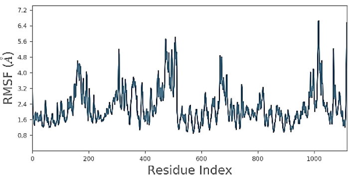
Figure 5: Root mean square fluctuation (RMSF). The figure shows the protein RMSF is moved 2.4 Å from residues between 1-100. then the movement decreases to 2a for 200-300 residues. Further, the protein then fluctuates to 5.6 Å for 500 residues, then sharply declines to 2.4 Å for residues till 1000. There is one sharp peak at the 1000 position with a decrease to 2.4 Å at the terminal. Please click here to view a larger version of this figure.

Figure 6: Protein secondary structure elements (SSE) and its residue index. The distribution of secondary structure elements (SSE) by residue index throughout the protein structure is shown in the plot above. The bottom plot tracks each residue's SSE assignment over time, whereas the plot below summarizes the SSE composition for each trajectory frame throughout the simulation. Please click here to view a larger version of this figure.
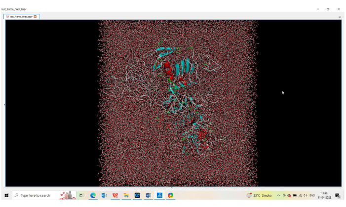
Figure 7: Simulations of lipid bilayers using the ENERGY force field. Simulations of lipid bilayers using the ENERGY force field. MD simulation is used to investigate geometrical features by showing the cartoon mode. Please click here to view a larger version of this figure.
Supplementary Figure 1: Addition of Kollman charges. Please click here to download this File.
Supplementary Figure 2: Minimizing pdb structure energy. Please click here to download this File.
Supplementary Figure 3: Preparing input files in Python. Please click here to download this File.
Supplementary Figure 4: Steps to create a Ramachandran plot. Please click here to download this File.
Supplementary Figure 5: Ligand creation. Please click here to download this File.
Supplementary Figure 6: Preparation of docking configuration. Please click here to download this File.
Supplementary Figure 7: Molecular dynamic simulation to create the docking complex. Please click here to download this File.
Supplementary Coding File 1. Please click here to download this File.
Supplementary Coding File 2. Please click here to download this File.
Supplementary Coding File 3. Please click here to download this File.
Supplementary Coding File 4. Please click here to download this File.
| タイプ | Total Num. | Concentration [mM] | Charge |
| Cl | 166 | 75.746 | -166 |
| Na | 111 | 50.65 | 111 |
Table 1: Counter ion/salt information.
Discussion
EGFR is the primary target receptor of breast cancer. EGFR overexpression increases breast cancer cases around the world. Meanwhile, specific antibodies such as single chain fragment variables are antibodies that move easily via blood circulation and have a fast clearance rate in the body. Antibodies are a wise solution and an effective immunotherapy drug37. Therefore, structure-based drug design must identify inhibitory medicines, such as scFv antibodies, that work specifically against a target receptor (EGFR). Based on in silico drug design, the investigator in this study worked to experiment with the stability of the scFv antibody-EGFR or protein-protein docked structure treated as a structure-based drug design. MD simulation methods were used in this study to investigate the stability and accuracy of the docked structure as a breast cancer inhibitor drug. Molecular biology experimental analysis mentioned in previous studies21, demonstrated the ability of scFv antibodies to bind with great affinity and specificity to EGFR.
This methodology is considered a successful approach to prove the stability of the binding interaction between the receptor and the antibody docking structure, which was mentioned in many investigations38,39. As shown in Figure 1A, D, in the structure's homology model software, writing the exact name of the files in the command sheet is the most critical step in that method. In addition, preparation of the 1ivo.pdb file is reasonably necessary to serve as a receptor without any external ligands that contaminate the docking process. In addition, Figure 1A,C shows Ramachandran plots for tool correction and accuracy detection of the homology model structure of the receptor and antibody structure models, and the plots show the numbers of residues in the α helices and β-sheet residues in the Ramachandran plot of the protein backbone of these structures. The more residues that exist in the α helices area of the β-sheet residues, the greater the accuracy of the structure. There are many ways to create a Ramachandran plot. Using www.ebi.ac.uk/thornton-srv/databases/pdbsum/ is a website that prepares the plot and shows the template names to show the residue positions.
Molecular docking is one of the most widely employed techniques in structure-based drug design. Protein-protein interactions require conformational rearrangements, particularly at the antibody-antigen interface. Such alterations may be as straightforward as the rotation of side chains or may involve more complicated changes, such as complete restructuring of the structure2. Therefore, specific technical limitations of this approach have been addressed, as docking works at a blind point of the grid box dimensions, and predicting the type and strength of the signal produced in the interaction process is always critical. Docking can predict the binding conformation of the ligands as a novel finding. The docking software used here is flexible, allowing ligands and receptors to interact with many possibilities when making grid boxes. As shown in Figures 2A,B, the lowest binding access was counted, and the connected residues are shown in Figure 2A.
Additionally, eight hydrogen bonds were recorded, as shown in Figure 3B, within 70 Å. The RMSD was refined, and the highest binding affinity creates a strong hydrogen bond with Asn70, which is considered crucial for activity and engages in significant additional interactions with other receptor-binding residues40, as shown in Figure 3A,B. The Verlet cutoff approach terminated the nonbounded interactions, Coulomb (electrostatic potential), and Lennard Jones (van der Waals attractions) interactions at 10 Å. As many H bonds are found in the protein-ligand connection, protein conformations are better for the interaction strength, as mentioned in previous studies29.
In a similar study1,19, the investigators revealed that, despite all of the structures being reasonably good quality and having slight structural variation, most approaches work on some crystal structures in the same protein family and fail on others. This study anticipated better consistency across various protein conformations of the same sequence. It is interesting to note that quality indicators such as resolution, Cruickshank diffraction precision index, or unresolved residues cannot accurately predict the success or failure of a specific structure. Additionally, cryptic sites were examined in many of those studies. Even though the researcher sought the binding site downloaded from the protein data bank (pdb) website as a reasonable approach to address the supposed anti-COVID-19 activity hypothesis, pdb is the primary source of the binding sites mentioned in many studies1,29.
In this study, MD simulation explained the potential energy analysis, a structural fluctuation analysis, and coordinate stability analysis. The calculated movements for the monomer of the protein experiencing the greatest conformational change in its unbound (free) form match the structural alterations that have been empirically seen to occur upon binding41. MD simulations are calculated by solving the classical equations of motion. The RMSD uses molecular dynamics (MD) to simulate how atoms and molecules interact in a system over a specific period. The RMSD is commonly calculated for the initial state of the structure. The deviation showed the most mobile parts of the structure42. In addition, the RMSD analyzes how much a structure differs from a reference over time. This simulation usually detected the stability of the docked structure. The RMSD was used to determine the conformation of the protein's strength and its deviation values produced during the simulation2,5,40,43. In the resulting numbers, the smaller the deviations, the more stable the protein structure is created. As shown in Figure 4, the RMSD value for the docked structure was recorded to be 20 ns-75 ns when the movement of the structure reached 7.5 Å, which proved the stability of the structure44.
However, RMSF calculates the RMSD average based on the time of the simulated structure45. High mobility is indicated by a region of the structure with high RMSF values frequently deviating from the reference structure, such as the receptor before stimulation. As shown in Figure 5, proteins are often only subjected to the backbone or α-carbon atom in RMSF. Therefore, RMSF analysis suggests conformational changes and shows more flexible side chains.
Finally, MD simulation was used to investigate geometrical features. By showing the cartoon mode in Figure 7, the membrane of EGFR protein topology and its movement are analyzed using RMSD as it differs from 20 ns-75 ns. The figure shows the protein sequence, with water and lipid molecules lined up and the phosphorus atoms of the lipids in beads15,46.
Even though protein-protein interactions are linked to important physiological processes and pathways, they are prospective targets for drug discovery. Small-molecule drugs, however, have not lived up to this promise. Many protein-protein interaction surfaces cannot accommodate the binding of small drug-like molecules as a result. Additionally, this study performed homology modeling, docking, and molecular dynamic simulations. The outcomes show that the scFv-antibody-receptor complex projected model exhibits excellent quality and lacks significant steric conflicts. As a result, this model might open the door for additional structural, functional, and therapeutic research that is specifically focused on in silico studies. Additionally, this research can lead to future drug-targeted studies for determining the procedures of different amino acid residues and predicting how receptor-antibody interactions and their motion change receptors, thereby affecting drug structure design. Thus, a geometric fit is only guaranteed following the structural reorganization of the proteins brought on by their recognition interaction.
開示
The authors have nothing to disclose.
Acknowledgements
None.
Materials
| Autodock software | Center for Computational structural Biology | AutoDock (scripps.edu) | |
| Desmond Maestro 19.4 software | Schrodinger | www.schrodinger.com | |
| Download Discovery Studio 2021 | Dassault Systems | https://discover.3ds.com/discovery-studio-visualizer-download. | |
| Modeler Version 9.24[17] | University of California | https://salilab.org/modeller/9.24/release.html | |
| Pictorial database of 3D structures (pdbsum) | EMBL-EBI | www.ebi.ac.uk/thornton-srv/databases/pdbsum/ | |
| PyMOL software | Schrodinger | PyMOL | pymol.org | |
| Pyrx software | Sourceforge | Download PyRx – Virtual Screening Tool (sourceforge.net) | |
| Python script 3.7.9 shell from the window (64) | Python | Python Release Python 3.7.9 | Python.org | |
| SPDBV software | Expasy | http://spdbv.vital-it.ch/disclaim.html |
参考文献
- Clark, J. J., Orban, Z. J., Carlson, H. A. Predicting binding sites from unbound versus bound protein structures. Sci Rep. 10 (1), 15856 (2020).
- Huang, Y., et al. A stepwise docking molecular dynamics approach for simulating antibody recognition with substantial conformational changes. Comput Struct Biotechnol J. 20, 710-720 (2022).
- Mahgoub, I. O., Ali, A. M., Hamid, M., Alitheen, N. M. Single chain fragment variable (scFv) secondary structure prediction and evaluation. FASEB J. 25, (2011).
- Kim, H., et al. Titanium dioxide nanoparticles induce apoptosis by interfering with EGFR signaling in human breast cancer cells. Environ Res. 175, 117-123 (2019).
- Mahgoub, E. O., Abdella, G. M. Improved exosome isolation methods from non-small lung cancer cells (NC1975) and their characterization using morphological and surface protein biomarker methods. J Cancer Res Clin Oncol. 149 (10), 7505-7514 (2023).
- Shaurova, T., Zhang, L., Goodrich, D. W., Hershberger, P. A. Understanding lineage plasticity as a path to targeted therapy failure in EGFR-mutant non-small cell lung cancer. Front Genet. 11, 281 (2020).
- Mahgoub, E. O., Bolad, A. K. Construction, expression and characterisation of a single chain variable fragment in the Escherichia coli periplasmic that recognise MCF-7 breast cancer cell line. J Cancer Res Ther. 10 (2), 265-273 (2014).
- Mahgoub, I. O. Design, expression and characterization of a single chain fragment variable anti-mcf-7 antibody; A humanized antibody derived from monoclonal antibody. Ann Res Conf Proc. 2014 (1), (2014).
- Sun, X. L., et al. Dimeric (-)-epigallocatechin-3-gallate inhibits the proliferation of lung cancer cells by inhibiting the EGFR signaling pathway. Chem Biol Interact. 365, 110084 (2022).
- Mahgoub, E. O., Haik, Y., Qadri, S. Comparison study of exosomes molecules driven from (NCI1975) NSCLC cell culture supernatant isolation and characterization techniques. FASEB J. 33, 647 (2019).
- Madeddu, C., et al. EGFR-mutated non-small cell lung cancer and resistance to immunotherapy: Role of the tumor microenvironment. Int J Mol Sci. 23 (12), 6489 (2022).
- Mahgoub, I., Bolad, A. K., Mergani, M. and immune-characterization of single chain fragment variable (scFv) antibody recognize breast cancer cells line (MCF-7). J. Immunother Cancer. 2, 6 (2014).
- Ladner, R. C., Sato, A. K., Gorzelany, J., de Souza, M. Phage display-derived peptides as therapeutic alternatives to antibodies. Drug Discov Today. 9 (12), 525-529 (2004).
- Khan, M. H., Manoj, K., Pramod, S. Reproductive disorders in dairy cattle under semi-intensive system of rearing in North-Eastern India. Vet World. 9 (5), 512-518 (2016).
- Al-Refaei, M. A., Makki, R. M., Ali, H. M. Structure prediction of transferrin receptor protein 1 (TfR1) by homology modelling, docking, and molecular dynamics simulation studies. Heliyon. 6 (1), 03221 (2020).
- Anbuselvam, M., et al. Structure-based virtual screening, pharmacokinetic prediction, molecular dynamics studies for the identification of novel EGFR inhibitors in breast cancer. J Biomol Struct Dyn. 39 (12), 4462-4471 (2021).
- Oduselu, G. O., Ajani, O. O., Ajamma, Y. U., Brors, B., Adebiyi, E. Homology modelling and molecular docking studies of selected substituted benzo d imidazol-1-yl) methyl) benzimidamide scaffolds on Plasmodium falciparum adenylosuccinate lyase receptor. Bioinform Biol Insights. 13, 1177932219865533 (2019).
- Mahgoub, E. O., et al. et al. of exosome isolation techniques in lung cancer. Mol Biol Rep. 47 (9), 7229-7251 (2020).
- Shree, P. Targeting COVID-19 (SARS-CoV-2) main protease through active phytochemicals of ayurvedic medicinal plants -Withania somnifera (Ashwagandha), Tinospora cordifolia (Giloy) and Ocimum sanctum (Tulsi) – a molecular docking study. J Biomol Struct Dyn. 40 (1), 190-203 (2022).
- Mahgoub, I. O. Expression and characterization of a functional single-chain variable fragment (scFv) protein recognizing MCF7 breast cancer cells in E. coli cytoplasm. Biochem Genet. 50 (7-8), 625-641 (2012).
- Mahgoub, E. O. Single chain fragment variables antibody binding to EGF receptor in the surface of MCF7 breast cancer cell line: Application and production review. Open J Genet. 7 (2), 84-103 (2017).
- Heng, C. K., Othman, R. Y. Bioinformatics in molecular immunology laboratories demonstrated: Modeling an anti-CMV scFv antibody. Bioinformation. 1 (4), 118-120 (2006).
- Mahgoub, E. O., Ahmed, B. Correctness and accuracy of template-based modeled single chain fragment variable (scFv) protein anti- breast cancer cell line (MCF-7). Open J Genet. 3 (3), 183-194 (2013).
- Khare, N., et al. Homology modelling, molecular docking and molecular dynamics simulation studies of CALMH1 against secondary metabolites of Bauhinia variegata to treat Alzheimer’s disease. Brain Sci. 12 (6), 770 (2022).
- Hu, J., Rao, L., Fan, X., Zhang, G. Identification of ligand-binding residues using protein sequence profile alignment and query-specific support vector machine model. Anal Biochem. 604, 113799 (2020).
- Santos, L. H. S., Ferreira, R. S., Caffarena, E. R. Integrating molecular docking and molecular dynamics simulations. Methods Mol Biol. 2053, 13-34 (2019).
- Xu, D., Tsai, C. J., Nussinov, R. Hydrogen bonds and salt bridges across protein-protein interfaces. Protein Eng. 10 (9), 999-1012 (1997).
- Das, S., Chakrabarti, S. Classification and prediction of protein-protein interaction interface using machine learning algorithm. Sci Rep. 11 (1), 1761 (2021).
- Mahmoud, S. S. A., Elkaeed, E. B., Alsfouk, A. A., Abdelhafez, E. M. N. Molecular docking and dynamic simulation revealed the potential inhibitory activity of opioid compounds targeting the main protease of SARS-CoV-2. Biomed Res Int. 2022, 1672031 (2022).
- Villada, C., Ding, W., Bonk, A., Bauer, T. Simulation-assisted determination of the minimum melting temperature composition of MgCl2-KCl-NaCl salt mixture for next-generation molten salt thermal energy storage. Front. Energy Res. 10, 809663 (2022).
- Hog, S. E., Rjiba, A., Jelassi, J., Dorbez-Sridi, R. NaCl salt effect on water structure: a Monte Carlo simulation study. Phys Chem Liq. 60 (5), 767-777 (2022).
- Maruyama, Y., Igarashi, R., Ushiku, Y., Mitsutake, A. Analysis of protein folding simulation with moving root mean square deviation. J Chem Inf Model. 63 (5), 1529-1541 (2023).
- Winarski, K. L., et al. Vaccine-elicited antibody that neutralizes H5N1 influenza and variants binds the receptor site and polymorphic sites. Proc Natl Acad Sci U S A. 112 (30), 9346-9351 (2015).
- Awal, M. A., et al. Structural-guided identification of small molecule inhibitor of UHRF1 methyltransferase activity. Front Genet. 13, 928884 (2022).
- Sharma, P., Hu-Lieskovan, S., Wargo, J. A., Ribas, A. adaptive, and acquired resistance to cancer immunotherapy. Cell. 168 (4), 707-723 (2017).
- Geerds, C., Wohlmann, J., Haas, A., Niemann, H. H. Structure of Rhodococcus equi virulence-associated protein B (VapB) reveals an eight-stranded antiparallel β-barrel consisting of two Greek-key motifs. Acta Crystallogr F Struct Biol Commun. 70 (7), 866-871 (2014).
- Mahgoub, E., et al. The therapeutic effects of tumor treating fields on cancer and noncancerous cells. Arabian J Chem. 14 (10), 103386 (2021).
- Hospital, A., Goni, J. R., Orozco, M., Gelpi, J. L. Molecular dynamics simulations: advances and applications. Adv Appl Bioinform Chem. 8, 37-47 (2015).
- Kaur, J., Tiwari, R., Kumar, A., Singh, N. Bioinformatic analysis of Leishmania donovani long-chain fatty acid-CoA ligase as a novel drug target. Mol Biol Int. 2011, 278051 (2011).
- Páll, S., Hess, B. A flexible algorithm for calculating pair interactions on SIMD architectures. Comp Phy Comm. 184 (12), 2641-2650 (2013).
- Tobi, D., Bahar, I. Structural changes involved in protein binding correlate with intrinsic motions of proteins in the unbound state. Proc Natl Acad Sci U S A. 102 (52), 18908-18913 (2005).
- Sinha, S., Wang, S. M. of VUS and unclassified variants in BRCA1 BRCT repeats by molecular dynamics simulation. Comput Struct Biotechnol J. 18, 723-736 (2020).
- Patel, D., Athar, M., Jha, P. C. Exploring Ruthenium-based organometallic inhibitors against Plasmodium falciparum calcium dependent kinase 2 (PfCDPK2): A combined ensemble docking, QM/MM and molecular dynamics study. Chem Select. 6 (32), 8189-8199 (2021).
- Khan, A., et al. Higher infectivity of the SARS-CoV-2 new variants is associated with K417N/T, E484K, and N501Y mutants: an insight from structural data. J Cell Physiol. 236 (10), 7047-7057 (2021).
- Zhang, Q., Shao, d., Xu, P., Jiang, Z. Effects of an electric field on the conformational transition of the protein: Pulsed and oscillating electric fields with different frequencies. Polymers. 14 (1), 123 (2021).
- Mahtarin, R., et al. et al. and dynamics of membrane protein in SARS-CoV-2. Biomol Struct Dyn. 40 (10), 4725-4738 (2022).

.
