Generating Murine Model of Myocardial Infarction: A Surgical Procedure for Permanent Ligation of Left Anterior Descending Coronary Artery in Mouse Model
Abstract
Source: Lugrin, J. et al. Murine Myocardial Infarction Model using Permanent Ligation of Left Anterior Descending Coronary Artery. J. Vis. Exp. (2019)
In this video, we describe a surgical procedure to generate a mouse model of myocardial infarction by permanently ligating the left anterior descending coronary artery.
Protocol
All procedures involving animal models have been reviewed by the local institutional animal care committee and the JoVE veterinary review board.
1. Anesthesia and Tracheal Cannulation
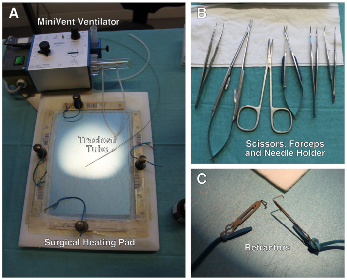
- Weigh the mouse to determine the dosage of anesthetic drugs, post-operative analgesic medication and tidal volume of the ventilator. Pre-warm the heating pad at 37 °C. The surgical setup is depicted in Figure 1.
- Inject mouse intraperitoneally with a mix of ketamine and xylazine at a dose of 80 mg/kg and 10 mg/kg, respectively.
- Quickly shave the mouse fur on the throat and the left side of the rib cage using an electric razor.
- Check depth of anesthesia by pinching tail and/or hind feet and settle the animal in a supine position on the heating pad. Place a small gauze compress under the head of the animal to avoid overheating of the eyes. Apply ocular gel to avoid eye dryness.
- Secure the four limbs with adhesive tape on the surface of the heating pad. Pass a loop of 5-0 silk suture under the upper incisors and stick the extremity of the loop with adhesive tape onto the heating pad. This will keep the mouth of the animal open and facilitate cannulation.
- Apply hair removal cream on the pre-shaved areas and gently massage with a cotton swab for 1 min. Wipe the excess of fur and cream with a gauze. Use drops of 0.9% saline solution and gauze to clean the incision areas. Apply pieces of sterile gauze to the shaved throat and thorax and soak them in iodopovidone.
NOTE: We recommend application of a local anesthetic drug (lidocaine or bupivacain) to incisions sites. - Set the ventilator at a tidal volume of 7 mL/kg and ventilation rate of 140 strokes/min.
NOTE: From now on, work under a microsurgery stereomicroscope. - Hold the skin on the center of the throat and perform an incision of 0.5 cm following a caudal/cephalic line using small scissors. Separate the lobes of salivary gland, then gently separate fascia of sternohyoid muscle with curved dissecting forceps until larynx and trachea are visible. Secure edges of the opening with retractors attached to elastic bands.
NOTE: Do this step without incision of the muscles. A trained operator will be able to intubate the animal via oral cavity without visualization of the trachea making this step optional. - Hold gently the tongue sideways. With forceps, insert the blunted inner needle of a 16 G cannula into the trachea. Visualize correct insertion into the trachea through the throat incision.
- Connect the cannula to the ventilator and ensure correct ventilation by placing the exhaust tubing into water. The presence of bubbles indicates correct intubation.
NOTE: In order to keep tissues wet during operation, place sterile gauze soaked with 0.9% saline solution and iodopovidone on the throat incision. Control moisture during the procedure.
2. Ligation of LAD Coronary Artery
- Release left anterior paw from duct tape and carefully move the mouse to right side decubitus position. Secure the left anterior limb once animal is in the correct position.
- Identify the line between left pectoralis minor and major muscles and make an oblique skin incision on 1 cm with scissors following the line. With dissecting blunt micro scissors, separate fascia of pectoralis muscles without incision. Maintain pectoralis muscles separated with retractors attached to elastic bands.
- Set the ventilator with a positive end-expiratory pressure (PEEP) of 3 cm H2O.
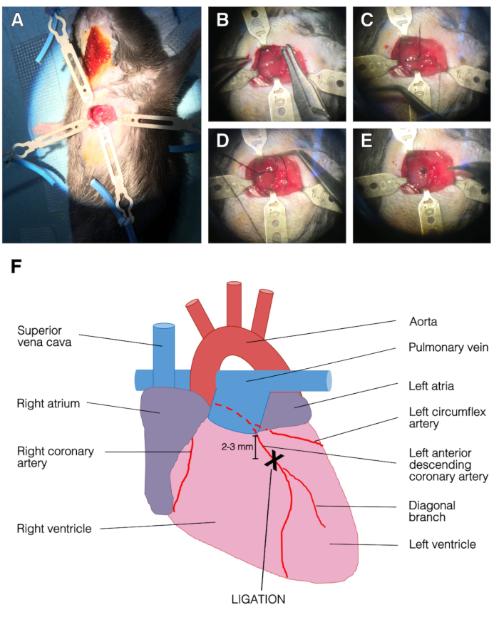
- Open the chest cavity by using blunt forceps at the 3rd intercostal space between 3rd and 4th ribs. Avoid touching internal thoracic artery as there is danger of bleeding. Do not touch heart or lung. Apply two retractors into the ribcage, one on each rib (Figure 2A).
- With a curved fine forceps, carefully remove the pericardium and pull it apart without harming the heart and lungs.
- Locate left anterior descending (LAD) coronary artery. LAD artery appears as a superficial bright red line running from the edge of the left auricle toward the apex.
- Use a needle holder to pass a 7-0 silk suture under the LAD 2 to 3 mm below left atria. Pull the silk slowly to avoid a tearing of heart tissue. Tie the ligature with three knots. The lower left part of the left ventricle will instantly turn pale upon ligation (Figure 2B–E).
NOTE: It is important to not go too deep into the ventricular cavity or to stay too superficial. For sham-operated animals, pull the suture silk under the LAD and remove it slowly avoiding tissue tearing. - Release the rib retractors, hold the 3rd rib with forceps and make two passes with a 6-0 silk suture under the 3rd and 4th ribs.
CAUTION: Do not the perforate heart or lung. Do not tighten knots yet. - Put three drops of 37 °C 0.9% saline solution onto the opening and shut the expiration exhaust tube for 2 or 3 respiratory cycles to properly inflate lungs. Tighten the suture and secure with two throws.
- Release retractors holding muscles and help them retrieve their correct place.
- Close thoracic skin with two stitches of 5-0 suture silk and secure with two throws. Close throat skin with one stitch of 5-0 suture silk and secure with two throws.
Representative Results

Figure 1: Description of the surgical setup. (A) Surgical setup comprises a modified heating pad, a ventilator and retractors attached to elastic bands. (B) Set of scissors, forceps and needle holder used during the surgery. (C) Close-up of the mini-retractors. Not shown: surgical stereomicroscope.

Figure 2: Representative images of the surgery and LAD ligation. (A) Opened chest with retractors. The left ventricle was apparent. Top, left and bottom retractors held the ribcage and right retractor held the pectoralis muscle. (B) The needle was passed under the LAD. (C) Suture silk was passed under the LAD, into the left ventricle. (D) Single stitch on the LAD. (E) End of the ligation procedure, the suture was secured with three knots. (F) Representation of an anterior view of the heart. The position of LAD ligation was 2-3 mm below left atria and above diagonal branch of the LAD.
開示
The authors have nothing to disclose.
Materials
| 30G Needle | BD Microlance 3 | 304000 | |
| Blunt Retractors | Fine Science Tools | 18200-09 | |
| Cotton Swabs | Applimed SA | 6001109 | |
| Dissecting Scissors, Curved | Aesculap | BC603R | |
| Electrical Razor | Remington | HC720 | |
| Hair Removal Cream, Veet | Silk & Fresh Tech. | 8218535 | |
| Micro Scissors, Curved Blunt/Blunt | Aesculap | FM013R | |
| 1 CC Syringe, Omnifix-F | B. Braun | 9161406V | |
| Silk Suture 5-0, BB | Ethicon | K880H | |
| Silk Suture 6-0, P-1 | Ethicon | 639H | |
| Silk Suture 7-0, BV-1 | Ethicon | K804 | |
| Student Dumont #7 Forceps | Fine Science Tools | 91197-00 | |
| Student Fine Forceps, Angled | Fine Science Tools | 91110-10 | |
| Surgical Gloves | Weitacare | 834301 | |
| Surgical heating pad | Personalized setting | ||
| Temgesic sol 0.3 mg/mL Buprenorphine | Indivior Schweiz AG | N02AE01 | |
| Tracheal tube inner needle of an 16G i.v. cat | Abbocath-T | G714-A01 | |
| Universal S3 Microscope, OMPIMD | Zeizz | ||
| Ventilator, MiniVent Model 845 | Harvard Apparatus | 73-0043 | |
| Viscotears | Alcon | 1551535 | |
| 70% Ethanol | |||
| Betadine 60 mL | MundiPharma |

