Summary

Handcrafted Silicone Coated Filament for Mice Middle Cerebral Artery Occlusion Models

Published: August 09, 2024
doi:

Summary

This protocol describes a straightforward method for creating coated filaments for the middle cerebral artery occlusion (MCAO) model in mice using silicone, nylon sutures, and syringe needles. This method allows for the production of filaments with a consistent diameter and various silicone wrapping lengths tailored to experimental needs.

Abstract

As the global population ages, ischemic stroke has risen to become the second leading cause of disability and mortality worldwide, placing an immense burden on both society and families. Although treatments such as intravenous thrombolysis and endovascular interventions can substantially improve the outcomes for patients with acute ischemic stroke, only a small percentage of individuals benefit from these therapies. To advance our understanding of the disease and to discover more effective treatments, researchers are continuously developing and refining animal models. Among these, the middle cerebral artery occlusion (MCAO) model stands out as the most commonly used model in cerebrovascular disease research. The filament used in this model is crucial for its development. This protocol outlines a method for creating filaments with consistent diameters and varying lengths of silicone coating. The MCAO model produced using this method in C57 mice has demonstrated high success and consistency, offering a valuable tool for tailored investigations into ischemic cerebrovascular diseases.

Introduction

Stroke is one of the most prevalent causes of mortality and disability worldwide. Ischemic and hemorrhagic strokes are the primary types of cerebrovascular event, with ischemic strokes accounting for approximately 87% of cases1,2,3. Currently, there are two treatment modalities for patients with ischemic stroke: pharmacological therapy with recombinant tissue plasminogen activator (rtPA) and mechanical thrombectomy. However, the narrow therapeutic window and extensive exclusion criteria limit the application of these treatments, benefiting only a minority of patients. This underscores the need for continued efforts to improve ischemic stroke therapies4,5. In vitro models are inadequate for replicating the complex pathophysiological responses following a stroke, making animal models an indispensable component of preclinical stroke research. Human focal cerebral ischemia is most frequently caused by thrombotic or embolic occlusion of the middle cerebral artery (MCA), which makes rodent models designed to simulate MCA occlusion (MCAO) highly relevant6.

The filament-induced MCAO model, the most widely adopted in stroke research, facilitates occlusion at the onset of the middle cerebral artery (MCA) and subsequent reperfusion, leading to extensive infarctions across subcortical and cortical areas of the brain. The advantage of this model lies in its ability to restore blood flow after inducing focal ischemia, thereby paralleling the pathophysiological processes observed in human stroke7. Additionally, the model simulates reperfusion injury, a critical factor in the extent of damage8. However, the MCAO model has limitations, including variability in infarct volume, with the standard deviation potentially reaching up to 64% of the mean value in some studies9. Despite over three decades of use, efforts to enhance the model's reliability are ongoing, yet significant variations in ischemic lesion volume persist across studies and laboratories10,11,12.

This article introduces a self-manufactured filament for inducing models evaluating neurological deficit scores and cerebral infarction areas. It examines the correlation between filament lengths coated with silicone and the success and stability of the MCAO model. This production technique yields filaments with commendable consistency, contributing to the development of a relatively stable MCAO model.

Protocol

All animal procedures adhered to the experimental procedures and standards approved by the Shanxi Provincial People's Hospital Institutional Animal Care and Use Committee (approval number: 2024 Provincial Medical Ethics Committee No. 64). The mice used in this experiment were male C57BL/6 mice, 8-10 weeks old, weighing 24-26 g. Details of the reagents and equipment used are listed in the Table of Materials.

1. Filament preparation

  1. Marking the original filament: Wind the 6-0 nylon suture evenly around a plastic ruler plate. Make marks at 5 mm and 10 mm from the filament head (including the coating mark point and the insertion depth mark point).
  2. Cut vertically downwards with a blade to ensure both ends are perfectly circular, resulting in an initial 2 cm long filament (Figure 1).
  3. Fabrication of the coating device: Use hemostatic forceps to snap off the needle head of a 26 G syringe, then polish the needle hole into a perfect circle with sandpaper. Draw up 2 mL of K-704 silicone sealant with a 10 mK syringe, and finally, attach the needle head to the syringe.
  4. Coating the filament: Insert the initial filament into the prepared needle hole up to the marked 5 mm or 10 mm position. Slowly and steadily push the syringe until the filament is fully coated under a stereomicroscope (Figure 2).
  5. Setting the coated filament: Fix the coated filament upright with adhesive tape and wait about 20 min for the silicone to fully set.
  6. Sterilization and packaging: Soak the prepared filaments in 75% alcohol, wipe them dry with a cotton swab, and then package them in 5 mL centrifuge tubes.

2. MCAO model

NOTE: Surgical tools were sterilized by autoclaving (121 °C at 15 psi for 60 min). The surgery table and other equipment were sanitized using 75% ethanol. The mice were fasted for 8 h preoperatively but allowed free access to water.

  1. Administer 5 mg/kg of meloxicam subcutaneously for analgesia 60 min prior to surgery. Connect a heat blanket to maintain the mouse's body temperature at 37 °C during anesthesia.
  2. Induce anesthesia with 4% isoflurane until spontaneous movements and whisker twitching cease, then maintain anesthesia at 1.5% (following institutionally approved protocols). Apply eye ointment to both eyes.
  3. Place the mouse in a supine position, secure its head and limbs, shave the hair on its neck and upper chest, and disinfect the skin with 75% ethanol from the inside out.
  4. Make a 2.5 cm long skin incision along the midline of the neck, from the lower jaw to the sternum.
  5. Bluntly dissect the right neck muscles to expose the carotid sheath. Use ophthalmic forceps to open the sheath and separate the common carotid artery (CCA), external carotid artery (ECA), and internal carotid artery (ICA), being careful to avoid disturbing the vagus nerve.
  6. Temporarily ligate the CCA with a slipknot before the bifurcation and clamp the ICA with a microsurgical artery clamp.
  7. Cauterize the superior thyroid artery from the ECA using a bipolar coagulation pen.
  8. Leave two threads on the ECA for ligation: one on the distal end for permanent ligation and another on the proximal end with a loose knot for future use. Make an approximately 0.5 mm incision between the two ligatures on the ECA using ophthalmic scissors to insert the filament.
  9. Insert the 5 mm or 10 mm silicone-coated filament into the CCA through the incision and then secure it by tightening the loose knot.
  10. After cutting off the distal end of the ECA and removing the clamp from the ICA, retract the filament to the CCA bifurcation. Then, flip and advance the filament into the deep ICA until one feels resistance. Slightly withdraw the filament and secure it by tightening the knot.
  11. Suture the animal's skin with 3-0 suture and disinfect the wound with iodine. Place the mouse in a recovery chamber for 1 h.
  12. Anesthetize the mouse again, gently remove the filament, tie off the ECA ligation thread securing the filament, and release the CCA slipknot to restore blood flow and reperfuse the middle cerebral artery.
  13. Trim the excess threads, suture the neck skin, and disinfect the area once more.

3. Sham operation

  1. For sham operations, insert a 7 mm silicone-coated filament to occlude the right middle cerebral artery and then immediately withdraw it to allow for instant reperfusion.
    NOTE: The subsequent procedure is identical to that performed on animals undergoing cerebral ischemia.

4. Neuroscore

  1. Place experimental animals from each group in an open field and conduct behavioral postoperative scoring 4 h after cerebral ischemia reperfusion.
  2. For successful modeling, consider scores between 1 and 3. The assessment criteria are based on the Longa scoring method10, as detailed in Table 1.
  3. Assess neurological deficits according to the Modified Neurologic Severity Scores (mNSS)13, with evaluations carried out at 24 h and 72 h post-reperfusion (see Table 2).

5. Transcardiac perfusion

  1. Anesthetize the mouse with 1.5% pentobarbital sodium (following institutionally approved protocols). Place the mouse back in its cage and wait for 10 min. Then, pinch the mouse's toes to test for the absence of reflexes and ensure deep anesthesia.
  2. Position the mouse in a supine position on a foam stand and secure its limbs.
  3. Cut off the tip of a 25 G needle to blunt it, preventing puncture of the aortic wall. Connect the needle to a syringe filled with 20 mL of saline.
  4. Lift the fur of the thorax and use scissors to cut away the skin to expose the xiphoid process. Grasp the xiphoid process and cut horizontally below it to expose the diaphragm by opening the muscle layer. Carefully cut the diaphragm with scissors, avoiding damage to the heart.
  5. Cut along the outer side of the sternum to open the rib cage on both sides, flip the anterior wall of the thorax, and secure it with hemostats.
  6. Use a cotton swab to remove the fat at the base of the heart, exposing the root of the aorta.
  7. Secure the heart with forceps, insert the needle at the apex of the heart, and advance obliquely upward until the needle is visible through the aortic wall. Clamp the needle in place.
  8. Make a small cut in the right atrium to observe blood flow. Steadily perfuse saline with the syringe, watching for blood to exit the right atrium. Once the effluent is clear, stop the perfusion14.
  9. After perfusion, decapitate the mouse to harvest the brain15 and place it in a -20 °C freezer for further processing.

6. Infarct volume assessment by TTC staining

  1. Freeze the procured brain tissues rapidly in a -20 °C freezer for 20 min, then place them on a pre-chilled brain slicing mold and section them into 1 mm thick slices.
  2. Immerse the obtained brain sections in 2% TTC solution and incubate at 37 °C for 20 min.
  3. Immerse the brain slices in 4% paraformaldehyde overnight and take photographs the following day.
  4. Measure the infarcted area for each slice and the total brain area using ImageJ. Calculate the infarct volume ratio using the formula: Infarct Volume % = (Sum of infarcted areas / Sum of total brain areas) × 100%.

Representative Results

In the creation of the MCAO model, the primary tools used for fabricating the filaments and the finished filaments are shown in Figure 3. Following filament production, the MCAO model is established by inserting the filament through the external carotid artery, with the duration of the operation recorded. Successful modeling is defined by a Longa score of 1-3 4 h post-filament withdrawal. Body weight is monitored daily after the operation. Neurological deficits are evaluated using modified neurological severity scores (mNSS) at 24 h and 72 h post-operation. Ischemic damage is assessed through TTC staining.

In the 10 mm filament group, five out of ten mice experienced subarachnoid hemorrhage (SAH), with two of these cases being fatal. Consequently, all five affected mice were excluded from the study. In contrast, none of the mice in the 5 mm filament group experienced SAH. The modeling success rate was 100% in the 5 mm filament group, significantly higher compared to the 50% success rate observed in the 10 mm filament group.

Surgical time, defined as the duration from skin incision to filament insertion, was significantly shorter in the 5 mm filament group compared to the 10 mm filament group (614 s ± 49.15 s vs. 758 s ± 65.63 s, P < 0.01) (Figure 4A). Both filament groups exhibited a significant reduction in postoperative body weight compared to the sham operation group; however, no significant difference was observed between the two filament sizes (Figure 4B).

TTC staining confirmed cerebral infarction in the self-made filament-induced MCAO model (Figure 5). Three days post-surgery, the infarct volume was 21.48% ± 6.79% for the 5 mm group and 19.85% ± 7.01% for the 10 mm group, with no significant statistical difference between the two groups regarding infarct size. In each group of mice, there was variability in the area of cerebral infarction, with three out of five mice showing a larger infarct area (Figure 5B,C) and two displaying a smaller infarct area (Figure 5E,F). Quantification of the infarct volume is shown in Figure 5G.

Neurological function deficits were assessed by mNSS scores on days 1 and 3 post-MCAO, as shown in Figure 6. On both days, the 5 mm and 10 mm groups exhibited significantly increased mNSS scores compared to the sham operation group, with no significant difference observed between the mNSS scores of the 5 mm and 10 mm groups.

Figure 1
Figure 1: Filament construction and marking. (A) Winding the 6-0 nylon suture evenly around a plastic ruler plate. (B) Labeling and vertically slicing the wound to create a 2 cm filament. Please click here to view a larger version of this figure.

Figure 2
Figure 2: Coating the filament. Please click here to view a larger version of this figure.

Figure 3
Figure 3: Finished filaments. Images of the completed filaments after coating and marking. Please click here to view a larger version of this figure.

Figure 4
Figure 4: Surgical duration and mouse body weight. (A) Surgical duration. (B) Mouse body weight measured before surgery and three days postoperatively. Statistical differences were analyzed using ANOVA and Tukey post hoc test. Data are presented as mean ± SEM, n = 5, **P < 0.01. Please click here to view a larger version of this figure.

Figure 5
Figure 5: TTC-stained brain sections. Representative TTC-stained brain sections showing areas of healthy tissue (red) and ischemic injury (white) for Sham (A,D), 5 mm MCAO (B,E), and 10 mm MCAO (C,F) groups. Three out of five mice show a larger infarct area (B,C), and two display a smaller infarct area (E,F). (G) Quantification of the infarct volume. Statistical differences were analyzed using ANOVA and Tukey post hoc test. Data are presented as mean ± SEM, n = 5, **P < 0.01. Please click here to view a larger version of this figure.

Figure 6
Figure 6: Neurological scores post-MCAO. Neurological scores recorded on the 1st and 3rd day post-MCAO. Statistical differences were analyzed using ANOVA and Tukey post hoc test. Data are presented as mean ± SEM, n = 5, ****P < 0.001. Please click here to view a larger version of this figure.

Figure 7
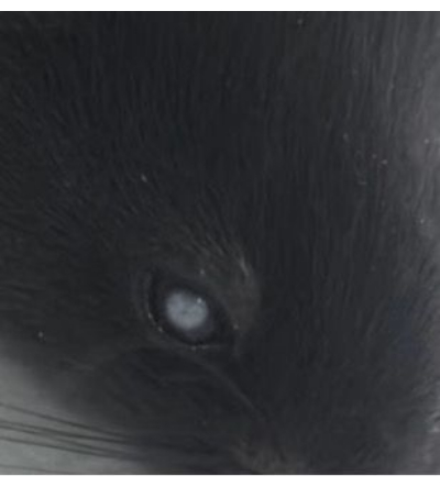
Figure 7: Pupil whitening post-filament insertion. Whitening of the pupil observed after insertion of the 10 mm silicone filament. Please click here to view a larger version of this figure.

0 points No symptoms of neurological damage
1 point Inability to fully extend the contralateral forelimb
2 points Circling to the contralateral side while walking
3 points Leaning to the contralateral side while walking
4 points Inability to walk spontaneously, loss of consciousness

Table 1: Longa scoring for model success. Longa scoring system used to determine the success of the model.

Tests Points
Raising mouse by tail 3
Flexion of forelimb 1
Flexion of hindlimb 1
Head moved >10° to vertical axis within 30s 1
Walking mouse on floor 3
Normal walk 0
Inability to walk straight  1
Circling toward the paretic side  2
Falling down to paretic side  3
Beam balance test 6
Balances with steady posture 0
Grasps side of the beam 1
Hugs the beam and 1 limb falls down from beam 2
Hugs the beam and 2 limbs fall down from beam, or spins on beam [>30 s] 3
Attempts to balance on beam but falls off [>20 s] 4
Attempts to balance on beam but falls off [>10 s] 5
Falls off, no attempt to balance or hang on the beam [<10 s] 6
Reflexes absence 2
Pinna reflex (a head shakes when touching the auditory meatus) 1
Corneal reflex (an eye blink when lightly touching the cornea with cotton) 1

Table 2: Modified Neurological Severity Scores (mNSS). Modified Neurological Severity Scores (mNSS) used to assess neurological severity.

Discussion

This study demonstrates a simple and cost-effective method for fabricating filament, confirming its feasibility in creating an MCAO model. The length of the filament's silicone coat can be adjusted according to experimental needs, offering additional flexibility. The preparation of a 5 mm filament embolus achieved a 100% success rate without any occurrences of subarachnoid hemorrhage (SAH) in mice. In the group using 10 mm filament emboli, there were instances of SAH, while the rest of the mice showed clear infarction in the MCA region. A critical step in reducing SAH was to slightly retract the filament upon encountering resistance during the procedure. In the preliminary experiment, nearly half of the mice without this adjustment experienced SAH when a 5 mm coated silicone filament was used. The filament encountered resistance upon reaching the mouse's anterior cerebral artery12. Therefore, any slight change in the mouse's position before withdrawing the filament may rupture the blood vessel. Retracting the filament slightly can effectively block the origin of the MCA while preventing the filament tip from entering the ACA and causing SAH.

When employing 10 mm filaments, a higher incidence of SAH was observed, which could be attributed to the reduced flexibility of the filament due to the increased silicone-coating length. This rigidity may cause the filament to puncture vessels when encountering resistance during insertion. Additionally, the surgical duration was longer for the 10 mm group, consistent with the increased difficulty of manipulating longer silicone-coated filaments, resulting in extended operation times. This indicates that shorter silicone-coated filament lengths can not only ensure a high rate of modeling success but also reduce surgical complexity and the occurrence of postoperative complications.

The finding that the use of 5 mm and 10 mm silicone-coated filaments in the MCAO model mice does not show significant differences in cerebral infarction areas and behavioral scores suggests that, under certain conditions, the length of the silicone coating on the filament may have minimal impact on experimental outcomes. However, within each group, there were noticeable differences in the cerebral infarction areas. In the 5 mm group, three out of five mice exhibited larger infarction areas, including regions such as the thalamus, hypothalamus, and hippocampus, while others had smaller infarction areas. The distribution of infarction sizes in the 10 mm group was similar to that of the 5 mm group, which may be related to the anatomical variability of the mice's cerebral vasculature15,16. A known contributing factor is the variation in the patency of the posterior communicating artery (PcomA)16. The PcomAs are the main collateral arteries following MCAO induced by the filament, supplying areas like the hippocampus and thalamus17. However, approximately 90% of C57BL/6 mice exhibit underdeveloped or absent PcomAs17, and the proportion of mice with larger infarction areas in each group is close to this percentage. Mice with underdeveloped or absent PcomAs, in addition to MCA, also have obstructed posterior cerebral artery (PCA) blood supply in the MCAO model, leading to larger infarction areas12. A study used shorter silicone-coated filaments (2 mm) to avoid obstructing the PCA blood supply, but this significantly reduced the model's success rate18. Variations in the anatomical distance between the MCA and the PCA at the juncture forming the Circle of Willis have been observed across different mouse strains and weight categories19. Consequently, to ensure modeling success and stability of infarct size within cohorts, it is critical to prepare silicone-coated filaments of tailored lengths that are adapted to the specific anatomical variations in the cerebral vasculature of mice across different strains and body weights. The method of filament production described in this article facilitates such research.

About 65% of ischemic stroke incidents are accompanied by temporary or permanent vision loss, usually attributed to affliction of the optic radiation or visual cortex20,21. However, stroke patients also carry a higher risk of acute retinal ischemia22,23. Currently, there are no clear experimental animal models of ischemic stroke that include retinal ischemia. In mice using 10 mm filaments, three mice experienced ipsilateral pupil whitening after filament insertion, which resolved about 5 min after the filament was withdrawn (Figure 7). This phenomenon indicates occlusion of the ophthalmic artery following filament insertion, but this was not observed in mice with 5 mm filaments. Currently, there is no consensus on the origin of the ophthalmic artery in experimental animals. Some studies suggest that, like in humans, the ophthalmic artery (OA) in C57BL/6J mice originates from the internal carotid artery24,25,26. Other research posits that, as in rats, the mouse OA originates from the pterygopalatine artery (PPA)27. Hence, considering the variability in the origin of the mouse OA, the pupil whitening observed in some mice from the 10 mm group could be due to this variability. In these mice, the ophthalmic arteries originating from the PPA experience insufficient blood supply due to occlusion at the start of the artery by the 10 mm silicone-coated filament upon insertion, resulting in pupil whitening. No such phenomenon occurred in the 5 mm group, likely because the shorter length of the silicone was insufficient to block the PPA.

Although this study provides an effective method for filament production and validates its efficacy experimentally, offering a strategy for the individualized preparation of filaments, it also has some limitations. The silicone coating on the handcrafted filaments is not uniformly applied, resulting in an irregular cylindrical shape. During the manufacturing process, the syringe must be operated at a constant speed to avoid inconsistencies in the silicone coating at the front and rear of the filament. Affordable tools and an easy-to-follow process make it feasible for beginners to practice the MCAO model with handcrafted filaments. Future research could explore improvements in filament design, such as optimizing materials or adjusting dimensions, to reduce the incidence of complications and enhance the reproducibility of the model. Moreover, accounting for anatomical differences to achieve a more stable model will be crucial for better understanding the mechanisms and treatment strategies of ischemic cerebrovascular diseases.

Divulgazioni

The authors have nothing to disclose.

Acknowledgements

This work was supported by the Wu Jieping Medical Foundation (320.6750.161290).

Materials

10 mL Syringe Haidike Medical Products Co., Ltd. Instrument for making filaments
2,3,5-Triphenyltetrazolium Chloride (TTC) Sigma-Aldrich G3005 Dye for TTC staining
24-well culture plate Corning  CLS3527 Vessel for TTC staining
26 G syringe needle Haidike Medical Products Co., Ltd. Instrument for making filaments
4% paraformaldehyde Servicebio G1101 Tissue fixation
6-0 nylon suture Haidike Medical Products Co., Ltd. Materials for making filaments
Anesthesia system for isoflurane Rwd Life Science Co., Ltd. R610  Anesthetized animal
Bipolar electrocoagulation generator Yirun Medical Instrument Co., Ltd. ZG300 Equipment for surgery
Constant temperature water bath Spring  Instrument Co., Ltd. HH-M6 TTC staining
Eye ointment Guangzhou Pharmaceutical H44023098 Material for surgery
Heat blanket ZH Biomedical Instrument Co., Ltd. Maintain body temperatur 
Isoflurane Rwd Life Science Co., Ltd. R510-22-10 Anesthetized animal
Meloxicam Boehringer-Ingelheim J20160020 Analgesia for animal
Microsurgical artery clamp Shanghai Jinzhong Surgical Instruments Co., Ltd.  W40130 Instrument for surgery
Microsurgical hemostatic clamp forceps Shanghai Jinzhong Surgical Instruments Co., Ltd.  M-W-0022 Instrument for surgery
Microsurgical instruments set Rwd Life Science Co., Ltd. SP0009-R Equipment for surgery
Mouse thermometer Hubei Dasjiaer Biotechnology FT3400 Intraoperative temperature monitoring
Pentobarbital sodium Sigma-Aldrich P3761 Euthanized animal
Shaver Joyu Electrical Appliances PHC-920 Equipment for surgery
Silicone Sealant Kafuter K-704 Materials for making filaments
Stereomicroscope Rwd Life Science Co., Ltd. 77001S Equipment for surgery
Suture thread with needle (3-0) Shanghai Pudong Jinhuan Medical Products Co., Ltd.  F404SUS302 Equipment for surgery

Riferimenti

  1. Collaborators, G. B. D. S. Global, regional, and national burden of stroke and its risk factors, 1990-2019: A systematic analysis for the global burden of disease study 2019. Lancet Neurol. 20 (10), 795-820 (2021).
  2. Kleindorfer, D. O. et al. 2021 Guideline for the prevention of stroke in patients with stroke and transient ischemic attack: A guideline from the American Heart Association/American Stroke Association. Stroke. 52 (7), e364-e467 (2021).
  3. Saini, V., Guada, L., Yavagal, D. R. Global epidemiology of stroke and access to acute ischemic stroke interventions. Neurology. 97 (20 Suppl 2), S6-S16 (2021).
  4. Hill, M. D. Coutts, S. B. Alteplase in acute ischaemic stroke: The need for speed. Lancet. 384 (9958), 1904-1906 (2014).
  5. Asif, K. S. et al. Mechanical thrombectomy global access for stroke (mt-glass): A mission thrombectomy (mt-2020 plus) study. Circulation. 147 (16), 1208-1220 (2023).
  6. Fluri, F., Schuhmann, M. K., Kleinschnitz, C. Animal models of ischemic stroke and their application in clinical research. Drug Des Devel Ther. 9, 3445-3454 (2015).
  7. Ringelstein, E. B. et al. Type and extent of hemispheric brain infarctions and clinical outcome in early and delayed middle cerebral artery recanalization. Neurology. 42 (2), 289-298 (1992).
  8. Shaik, N. F., Regan, R. F., Naik, U. P. Platelets as drivers of ischemia/reperfusion injury after stroke. Blood Adv. 5 (5), 1576-1584 (2021).
  9. Zhang, S. R. et al. Large-scale multivariate analysis to interrogate an animal model of stroke: Novel insights into poststroke pathology. Stroke. 52 (11), 3661-3669 (2021).
  10. Longa, E. Z., Weinstein, P. R., Carlson, S., Cummins, R. Reversible middle cerebral artery occlusion without craniectomy in rats. Stroke. 20 (1), 84-91 (1989).
  11. Chen, Y., Ito, A., Takai, K., Saito, N. Blocking pterygopalatine arterial blood flow decreases infarct volume variability in a mouse model of intraluminal suture middle cerebral artery occlusion. J Neurosci Methods. 174 (1), 18-24 (2008).
  12. Yuan, F. et al. Optimizing suture middle cerebral artery occlusion model in c57bl/6 mice circumvents posterior communicating artery dysplasia. J Neurotrauma. 29 (7), 1499-1505 (2012).
  13. Bieber, M. et al. Validity and reliability of neurological scores in mice exposed to middle cerebral artery occlusion. Stroke. 50 (10), 2875-2882 (2019).
  14. Gage, G. J., Kipke, D. R., Shain, W. Whole animal perfusion fixation for rodents. J Vis Exp. 65, e3564 (2012).
  15. Barone, F. C., Knudsen, D. J., Nelson, A. H., Feuerstein, G. Z., Willette, R. N. Mouse strain differences in susceptibility to cerebral ischemia are related to cerebral vascular anatomy. J Cereb Blood Flow Metab. 13 (4), 683-692 (1993).
  16. Kitagawa, K. et al. Cerebral ischemia after bilateral carotid artery occlusion and intraluminal suture occlusion in mice: Evaluation of the patency of the posterior communicating artery. J Cereb Blood Flow Metab. 18 (5), 570-579 (1998).
  17. Mccoll, B. W., Carswell, H. V., Mcculloch, J., Horsburgh, K. Extension of cerebral hypoperfusion and ischaemic pathology beyond mca territory after intraluminal filament occlusion in c57bl/6j mice. Brain Res. 997 (1), 15-23 (2004).
  18. Liu, Z. et al. Optimisation of a mouse model of cerebral ischemia-reperfusion to address issues of survival and model reproducibility and consistency. Comput Intell Neurosci. 2022, 7594969 (2022).
  19. Ward, R., Collins, R. L., Tanguay, G., Miceli, D. A quantitative study of cerebrovascular variation in inbred mice. J Anat. 173, 87-95 (1990).
  20. Hanna, K. L., Hepworth, L. R., Rowe, F. Screening methods for post-stroke visual impairment: A systematic review. Disabil Rehabil. 39 (25), 2531-2543 (2017).
  21. Rowe, F. J. Stroke survivors' views and experiences on impact of visual impairment. Brain Behav. 7 (9), e00778 (2017).
  22. Scoles, D., Mcgeehan, B., Vanderbeek, B. L. The association of stroke with central and branch retinal arterial occlusion. Eye (Lond). 36 (4), 835-843 (2022).
  23. Kim, Y. D. et al. Cerebral magnetic resonance imaging of coincidental infarction and small vessel disease in retinal artery occlusion. Sci Rep. 11 (1), 864 (2021).
  24. Emiroglu, M. Y. et al. Effects of obstructive carotid artery disease on ocular circulation and the safety of carotid artery stenting. Heart Lung Circ. 26 (10), 1069-1078 (2017).
  25. Cotofana, S. Lachman, N. Arteries of the face and their relevance for minimally invasive facial procedures: An anatomical review. Plast Reconstr Surg. 143 (2), 416-426 (2019).
  26. Xu, X. et al. Dibazol-induced relaxation of ophthalmic artery in C57bl/6J mice is correlated with the potency to inhibit voltage-gated ca(2+) channels. Exp Eye Res. 231, 109468 (2023).
  27. Justic, H. et al. Redefining the Koizumi model of mouse cerebral ischemia: A comparative longitudinal study of cerebral and retinal ischemia in the Koizumi and Longa middle cerebral artery occlusion models. J Cereb Blood Flow Metab. 42 (11), 2080-2094 (2022).
This article has been published
Video Coming Soon
Keep me updated:

.

Citazione di questo articolo
Zhang, M., Yuan, Y., Du, R., Wen, C., Lin, S., Zhang, Y., Xiang, Z., Hu, F., Wang, C. Handcrafted Silicone Coated Filament for Mice Middle Cerebral Artery Occlusion Models. J. Vis. Exp. (210), e67002, doi:10.3791/67002 (2024).

View Video