Salinity-dependent Toxicity Assay of Silver Nanocolloids Using Medaka Eggs
Summary
Embryonic stages are the most susceptible to xenobiotics. Although chemical toxicity depends on salinity, no method exists to test the salinity dependence of toxicity to aquatic organisms. Here, we describe a new and high-throughput method for determining the salinity dependence of toxicity to aquatic embryos.
Abstract
Salinity is an important characteristic of the aquatic environment. For aquatic organisms it defines the habitats of freshwater, brackish water, and seawater. Tests of the toxicity of chemicals and assessments of their ecological risks to aquatic organisms are frequently performed in freshwater, but the toxicity of chemicals to aquatic organisms depends on pH, temperature, and salinity. There is no method, however, for testing the salinity dependence of toxicity to aquatic organisms. Here, we used medaka (Oryzias latipes) because they can adapt to freshwater, brackish water, and seawater. Different concentrations of embryo-rearing medium (ERM) (1x, 5x, 10x, 15x, 20x, and 30x) were employed to test the toxicity of silver nanocolloidal particles (SNCs) to medaka eggs (1x ERM and 30x ERM have osmotic pressures equivalent to freshwater and seawater, respectively). In six-well plastic plates, 15 medaka eggs in triplicate were exposed to SNCs at 10 mg/L−1 in different concentrations of ERM at pH 7 and 25 °C in the dark.
We used a dissecting microscope and a micrometer to measure heart rate per 15 sec and eye diameter on day 6 and full body length of the larvae on hatching day (section 4). The embryos were observed until hatching or day 14; we then counted the hatching rate every day for 14 days (section 4). To see silver accumulation in embryos, we used inductively coupled plasma mass spectrometry to measure the silver concentration of test solutions (section 5) and dechorionated embryos (section 6).The toxicity of the SNCs to medaka embryos obviously increased with increasing salinity. This new method allows us to test the toxicity of chemicals in different salinities.
Introduction
Since the establishment of the Organisation for Economic Co-operation and Development (OECD) test guidelines for testing chemicals in 1979, 38 test guidelines have been published in Section 2 of the guidelines, Effects on Biotic Systems1. All of the aquatic organisms tested have been from freshwater habitats, namely freshwater plants; algae; invertebrates such as daphnia and chironomids; and fishes such as medaka, zebrafish, and rainbow trout. Compared to saltwater environments, freshwater environments are more directly affected by human economic and industrial activities. Therefore, freshwater environments have been prioritized for testing because they are at higher risk from pollution.
In coastal areas, including estuaries, salinities vary between brackish water and seawater conditions, and these areas are often polluted by industrial activity2. Coastal areas and their associated wetlands are characterized by high ecological biodiversity and productivity. Coastal ecosystems must therefore be protected from chemical pollution. However, there has been limited ecotoxicological research in brackish water and seawater habitats.
Sakaizumi3 studied the toxic interactions between methyl mercury and salinity in Japanese medaka eggs and found that increasing the osmotic pressure of the test solution enhanced the toxicity of the methyl mercury. Sumitani et al.4 used medaka eggs to investigate the toxicity of landfill leachate; they found that the osmotic equivalency of leachate to the eggs was the key to inducing abnormalities during embryogenesis. In addition, Kashiwada5 reported that plastic nanoparticles (39.4 nm in diameter) easily permeated through the medaka egg chorion under brackish conditions (15x embryo rearing medium (ERM)).
A typical small fish model, the Japanese medaka (Oryzias latipes) has been used in basic biology and ecotoxicology6. Japanese medaka can live in conditions ranging from freshwater to seawater because of their highly developed chloride cells7. They are therefore likely to be useful for testing in conditions with a wide range of salinities.
Protocol
The Japanese medaka used in this study were treated humanely in accordance with the institutional guidelines of Toyo University, with due consideration for the alleviation of distress and discomfort.
1. Silver Nanocolloids (SNCs)
- Purchase purified SNCs (20 mg/L−1, 99.99% purity, particle mean diameter about 28.4 ± 8.5 nm suspended in distilled water).
- Validate the purity and concentration of the silver by inductively coupled plasma mass spectrometry (ICP-MS) analyses according to operating manual8. The pretreatment method for ICP-MS analyses is described in section 7.
2. Preparation of SNC Solutions (Mixtures of Silver Colloids and Ag+) with Different Salinities
- Prepare 60× ERM consisting of 60 g NaCl, 1.8 g KCl, 2.4 g CaCl2·2H2O, and 9.78 g MgSO4·7H2O in 1 L of ultrapure water; adjust the pH to 7.0 with 1.25% NaHCO3 in ultrapure water.
- Stir the ERM solution at 25 °C overnight.
- Mix SNCs with diluted ERM. Prepare 40 ml of each SNC-ERM mixed solution. The final concentration is 10 mg/L−1 of SNCs in different concentrations of ERM (1x, 5x, 10x, 15x, 20x, or 30x).
- Adjust pH of the SNC-ERM mixed solution to 7.0 with 0.625% NaHCO3 in ultrapure water. pH adjustment is very important in preparing the SNC solution, because Ag+ release is facilitated by acidic conditions9.
- Use AgNO3 as a reference compound for SNCs.
- Mix AgNO3 with diluted ERM. Prepare 40 ml of AgNO3-ERM mixed solution at an AgNO3 concentration of 15.7 mg/L-1 (10 mg/L−1 silver) in different concentrations of ERM (1x, 5x, 10x, 15x, 20x, or 30x).
Note: To examine silver colloid toxicity, AgNO3 solution, which is a source of soluble silver, is used as a reference compound for SNCs, which are a mixture of silver colloids and soluble silver.
- Mix AgNO3 with diluted ERM. Prepare 40 ml of AgNO3-ERM mixed solution at an AgNO3 concentration of 15.7 mg/L-1 (10 mg/L−1 silver) in different concentrations of ERM (1x, 5x, 10x, 15x, 20x, or 30x).
3. Medaka Culture and Egg Harvesting
- Obtain the medaka (O. latipes) (orange-red strain) (60 males and 60 females).
- Culture medaka as groups (20 males and 20 females as one group) in 1x ERM in 3 L tanks by using a medaka flow-through culturing system.
- Culture at the following conditions:
pH range of the culture medium: 6.2 to 6.5
light:dark cycle: 16:8 hr
temperature of the culture medium: 24 ± 0.5 °C
osmotic pressure of the culture medium: 257 mOsm
- Culture at the following conditions:
- Feed medaka on Artemia salina nauplii at 10:00 (once a day) and feed an artificial dry fish diet at 09:00, 11:00, 13:00, 15:00, and 17:00 (five times a day).
- Obtain A. salina nauplii.
- Prepare 5 L of a 3.0% salt solution in a plastic beaker.
- Add 30 g of brine shrimp eggs to the salt solution in the beaker.
- Incubate the eggs at 25 °C for 48 hr with bubbling (4 L/min−1) using an aeration pump.
- After 48 hr, stop the bubbling.
- Allow the solution to stand for 5 to 10 min to separate the hatched A. salina nauplii (lower part of the solution) from the unhatched eggs and eggshells (upper part of the solution).
- Remove the upper layer of the solution by decantation.
- Filter the lower portion of the solution through a sieve with openings of 283 µm, and collect the nauplii that pass through on a net with openings of 198 µm.
- Feed the nauplii to the medaka within 6 hr.
- After the female medaka have spawned, remove the external egg clusters gently from the females' bodies or collect the eggs from the bottom of the fish tank by using a small net (net size 5 cm x 5 cm, hole size 0.2 mm x 0.2 mm).
- Rinse the egg cluster with flowing tap water for 5 sec.
- Add all of the rinsed egg clusters to 30x ERM solution.
- Remove the clusters from the solution after 1 min and place the egg clusters between dry paper towels and roll gently.
- Put the eggs back into the 30x ERM.
- Select fertilized eggs under a dissecting microscope.
- Place selected 810 eggs in 1x ERM in six-well plastic plates by using forceps.
- Incubate the eggs at 25 ± 0.1 °C in an incubator until developmental stage 21. (Developmental stages of the medaka embryos were defined from the work of Iwamatsu10.)
- Pick out incubated eggs at developmental stage 21 under a dissecting microscope.
- Rinse selected eggs with 1x ERM.
- Subject the rinsed eggs to exposure experiments (section 4).
4. Toxicity Testing of SNCs or AgNO3 at Different ERM Salinities
- Rinse medaka eggs (stage 21) three times with test solution [SNCs (10 mg/L−1) or AgNO3 (15.7 mg/L−1 as 10 mg/L−1 silver) at each concentration of ERM (1x, 5x, 10x, 15x, 20x, or 30x) at pH 7]. As controls, use eggs in 1× to 30× ERM at pH 7.
- Add 15 rinsed eggs to 5 ml of each test solution in six-well plastic plates. (Perform the exposure experiments three times for SNC or AgNO3 toxicity testing using each test solution.)
- Wrap the plates in aluminum foil.
- Incubate the wrapped plates at 25 °C in the dark until hatching or for 14 days.
- Observe the exposed eggs every 24 hr for biological changes and dead eggs (Figures 1 and 2).
- Exchange the test solutions every 24 hr.
- Perform observations as follows.
- On day 6 of exposure, count the heart rate (per 15 sec) of medaka embryos under a dissecting microscope by using a stopwatch (Figure 3a).
- On day 6 of exposure, measure the eye size (diameter) of medaka embryos under a dissecting microscope by using a micrometer (Figure 3b).
- On hatching day, measure the full body lengths of larvae under a dissecting microscope by using a micrometer (Figure 3c).
- Count the total number of exposed eggs that hatch over the 14 days (Figure 3d).
5. Isolation of Soluble Silver from SNC Solution, and Silver Analysis
- Isolate soluble silver from each SNC solution (a mixture of silver colloids and soluble silver) by filtering through a 3 kDa membrane filter at 14,000 x g and 4 °C for 10 min. Use a 3 kDa membrane filter to isolate soluble silver from the SNCs, because the reported mean diameter of aggregated SNCs in 1x ERM is 67.8 nm11 and that of Ag+ is 0.162 nm12; the 3 kDa membrane excludes particles with diameters of 2 nm or more13.
- Measure the silver concentration in 50 µl of filtered solution (= the soluble silver concentration) by ICP-MS analysis (Figure 3e) according to the ICP-MS operating manual8. The pretreatment method for the ICP-MS analyses is described in section 7.
6. Measurement of Silver Bioaccumulation in Medaka Embryos
- Expose medaka eggs (stage 21) to SNCs or AgNO3 as described in section 4.
- On day 6 of exposure, remove chorion from the egg (i.e., dechorion) by using medaka hatching enzyme according to the protocol described in the Medaka Book14.
- Measure the silver concentration of the dechorioned eggs by ICP-MS analysis according to the ICP-MS operating manual8 (Figure 3f). The pretreatment method for the ICP-MS analyses is described in section 7.
7. Measurement of Silver Concentration by ICP-MS Analysis
- Add samples [50 µl of silver solution (for validation of the silver concentration; section 1); three dechorionated embryos (section 5); or 50 µl of filtered solution (section 5)] to a 50 ml Teflon beaker.
- Add 2.0 ml of ultrapure nitric acid to the 50 ml beaker.
- Heat the mixture on a hot plate at 110 °C until just before it dries out (about 3 hr).
- To dissolve the organic matter completely, add 2.0 ml of ultrapure nitric acid and 0.5 ml of hydrogen peroxide to the beaker.
- Heat the mixture again on the hot plate until just before it dries out (about 3 hr).
- Dissolve the residue in 4 ml of 1.0% ultrapure nitric acid solution.
- Transfer 4 ml of solution to a centrifuge tube.
- Repeat 7.6 to 7.7 twice (a total of three times). The final volume is 12.0 ml.
- Measure the silver concentration of the sample (dissolved in 1.0% ultrapure nitric acid) by using ICP-MS analysis according to the operating manual8.
- Use an internal and an external standard solution (See Materials List) to quantify the silver concentration. The internal and external standard solution is accredited by American Association for Laboratory Accreditation (A2LA). Detection limits of silver were 0.0018 ng/ml−1 (solution) and 0.016 ng mg-weight−1 (embryo body).
Representative Results
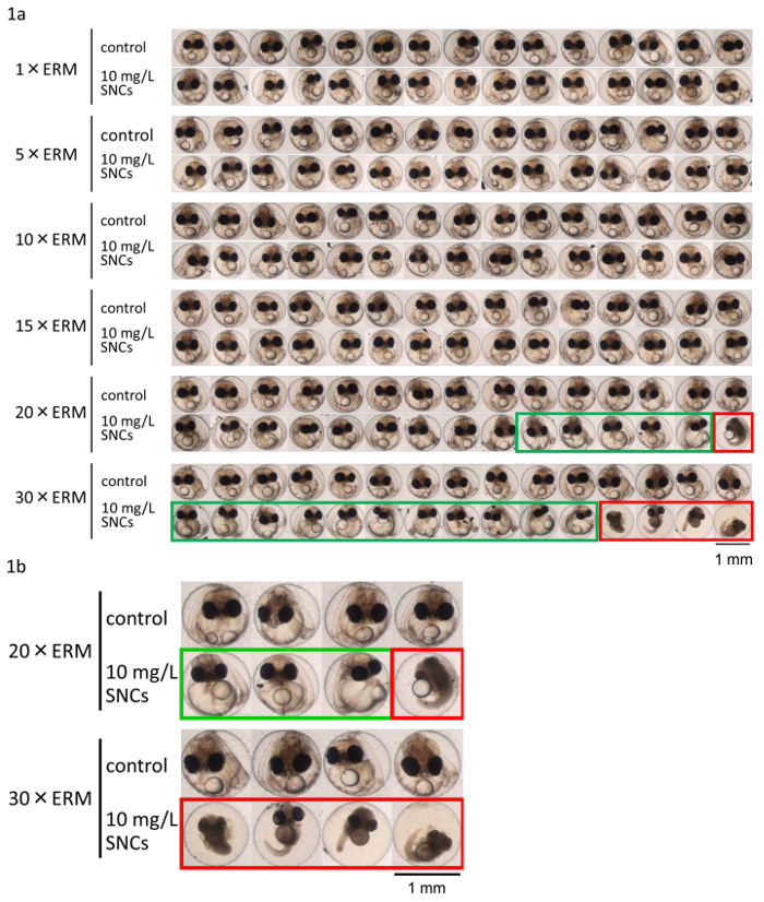
The effect of salinity on SNC toxicity was very obvious: the induction of deformity or death was salinity dependent (Figures 1 and 2). We measured phenotypic biomarkers (heart rate, eye size, full body length, and hatching rate) in SNC (10 mg/L−1)-exposed embryos. These phenotypic biomarkers revealed salinity-dependent SNC toxicity.
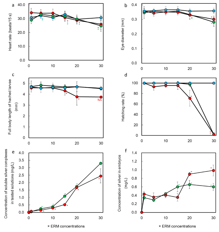
Heart rates ranged from 29.6 to 32.2 beats/15 sec throughout 1x to 30x ERM in the controls. However, they decreased significantly (P <0.01) with SNC or AgNO3 exposure in 30x ERM (Figure 3a). Decreasing heart rate indicates deteriorating health. There were no significant differences in full body length of the larvae under control or AgNO3 exposure at salinities ranging from 5x to 30x ERM compared with the respective 1x ERM solutions. Body length was consistently 4.55 to 4.69 mm. However, body length decreased significantly (P <0.01) to 4.33 and 3.77 mm, as a result of SNC exposure in 15x and 20x ERM compared with the respective 1x ERM solutions; moreover, it decreased to 3.75 mm in 30x ERM (statistical analysis was not available at 30x ERM because only one hatched) (Figure 3c). Decreasing full body length indicates growth inhibition. There were no significant differences in eye diameter in the controls at salinities ranging from 1x to 30x ERM compared with 1x ERM; eye diameter was consistently 0.357 to 0.366 mm. However, it decreased significantly upon SNC or AgNO3 exposure in 20x or 30x ERM compared with in the respective 1x ERM solutions (Figure 3b). Decreasing eye diameter indicates developmental inhibition of the nervous system. All control eggs hatched within 14 days. However, upon SNC exposure in 20x and 30x ERM the hatching rate decreased significantly to 71% and 2%, respectively, of the rate in 1x ERM (P <0.01) (Figure 3d). Also, upon AgNO3 exposure it decreased significantly in 30x ERM (P <0.01). Decreasing hatching rate indicates the toxic effect of the presence of SNCs or AgNO3. These four phenotypic biomarkers therefore show salinity dependent SNC toxicity.
Salinity increases water-soluble metal complex formation, and these complexes might have toxic effects3,8. In our study, ICP-MS analyses of silver revealed that the soluble silver concentrations in the test solutions increased as the salinity increased; the silver concentration in the embryos also increased (Figures 3e, 3f).

Figure 1: Increasing salinity increases SNC toxicity. Mortality and number of abnormally developed embryos increased with increasing salinity under SNC exposure. (a) Image array of medaka eggs exposed to 10 mg/L−1 SNC solution at different ERM concentrations. Images are typical of medaka eggs exposed to SNCs and observed under a dissecting microscope. Control medaka eggs were well developed, and all of them hatched in 1x to 30x ERM. At 10 mg/L−1 SNC exposure, although all of the medaka eggs hatched in 1x to 15x ERM, developmental deformities (red outlined rectangles, unhatched) and embryos unhatched within 14 days (green outlined rectangles, unhatched) were observed at 20x and 30x ERM. (b) Magnified images of the lower right of (a). Please click here to view a larger version of this figure.

Figure 2: Typical phenotypic biomarkers of medaka eggs exposed to SNCs. Medaka eggs at developmental stage 21 were exposed to SNCs (10 mg/L−1) in different concentrations of ERM for 6 days. (a) Control medaka embryo with normal development. (b) Developmental deformity (light degree of damage). This embryo displayed pericardiovascular edema; tubular heart; blood clots; inadequate development of the blood vessels (and thus ischemia), spinal cord, tail, eyes, and brain; and a short tail. (c) Developmental deformity (heavy degree of damage). This embryo showed destruction of the yolk sack; inadequate development of the blood vessels (and thus ischemia), spinal cord, tail, eyes, and brain; and a short tail. The signs in (b) and (c) were observed upon SNC exposure in 20x and 30x ERM. Please click here to view a larger version of this figure.

Figure 3: Effects of exposure to SNCs or silver nitrate on toxicological biomarkers during medaka egg development. Developmental stage 21 medaka eggs exposed to SNCs (10 mg/L−1) or silver nitrate (10 mg/L−1 as silver) in a series of ERMs were observed for 6 days. [blue] Control (ERM); [red] SNCs at 10 mg/L−1 in ERM; [green] AgNO3 at 10 mg/L−1 as silver in ERM. (a) Heart rate per 15 sec. Decreasing heart rate indicates deteriorating health. (b) Eye diameter. Decreasing eye diameter indicates developmental inhibition of the nervous system. (c) Full body length. Decreasing full body length indicates growth inhibition. (d) Hatching rate. Decreasing hatching rate indicates the toxic effect of the presence of SNCs. (e) Concentrations of soluble silver complexes from SNCs or silver nitrate in test solutions (mg/L−1). (f) Silver concentrations in embryos exposed to SNCs or silver nitrate in a series of ERMs. *Significant difference (analysis of variance, P <0.05) compared with the respective 1x ERM solution. NA: not available because only one hatched. Error bars indicate standard deviation. Please click here to view a larger version of this figure.
Discussion
Medaka is a freshwater fish that is highly tolerant to seawater; it is not well known that the original natural habitat of this fish was saltwater off the Japanese coast6. Hence, medaka fish have well-developed chloride cells7. This unique property provides scientists with a new way to test the toxicity of chemicals in the environment as a function of salinity (freshwater to seawater) by using only a single species of fish.
To obtain medaka eggs at stage 21, eggs must be harvested every morning and selected at stage 20. Usually, medaka pairs start mating in the early morning (just before sunrise) and produce eggs by sunrise. Eggs harvested in the morning must be at about stage 10 or 11. If there is a need to control egg development before the start of the experiment, egg development can be slowed by using temperatures of 15 to 20 °C before stage 21 is reached. Measuring the silver concentration (soluble silver) in the test solutions and in dechorionated embryos was important to our investigation of the salinity dependence of SNC toxicity. Hatching enzyme is the best biologically suitable enzyme for removing the chorion, because its high specificity means that it has no harmful proteinase. Other proteinases are not recommended. So far, the only hatching enzyme available is that for medaka; this is one limitation of this method.
The obvious effect of salinity on the outcome of the chemical toxicity tests demonstrated that simulating such natural aquatic properties as realistically as possible, as in our experiments, was useful for investigating the toxicity of chemicals in the environment. The discovery that SNC toxicity due to high silver concentrations was increased by salinity is highly applicable to the ecotoxicology of pollutant chemicals in all aquatic areas. In the case of general chemical toxicity testing in seawater, there is as yet no fish model nominated by authorized international organizations (e.g., the OECD and International Organization for Standardization). Among the freshwater fishes (e.g., medaka, zebrafish, carp, rainbow trout, and fathead minnow) that have been used for chemical toxicity testing, only the medaka has all of the advantages of salinity adaptation, hatching enzyme availability, high fecundity, and a size sufficiently small for easy use in laboratory experiments. Furthermore, medaka can be adapted to a wide temperature range (2 to 38 °C)6. In aquatic environments, salinity and temperature are the most important environmental influences on the fate of chemicals; our method should therefore be modifiable for a range of aquatic environmental research.
Divulgations
The authors have nothing to disclose.
Acknowledgements
We are grateful to Ms. Kaori Shimizu and Mr. Masaki Takasu of the Graduate School of Life Sciences, Toyo University, for their technical support. This project was supported by research grants from the Special Research Foundation and Bio-Nano Electronics Research Centre of Toyo University (to SK); by the Science Research Promotion Fund of the Promotion and Mutual Aid Corporation for Private Schools of Japan (to SK); by the New Project Fund for Risk Assessments, from the Ministry of Economy, Trade and Industry (to SK); by a Grant-in-Aid for Challenging Exploratory Research (award 23651028 to SK); by a Grant-in-Aid for Scientific Research (B) and (C) (award 23310026 and 26340030 to SK); and by a Grant-in-Aid for Strategic Research Base Project for Private Universities (award S1411016 to SK) from the Ministry of Education, Culture, Sports, Science and Technology of Japan.
Materials
| Silver nanocolloids | Utopia Silver Supplements | ||
| NaCl | Nacalai Tesque, Inc. | 31319-45 | For making ERM |
| KCl | Nacalai Tesque, Inc. | 28513-85 | For making ERM |
| CaCl2·2H2O | Nacalai Tesque, Inc. | 06730-15 | For making ERM |
| MgSO4·7H2O | Nacalai Tesque, Inc. | 21002-85 | For making ERM |
| NaHCO3 | Nacalai Tesque, Inc. | 31212-25 | For making ERM |
| AgNO3 | Nacalai Tesque, Inc. | 31018-72 | |
| pH meter | HORIBA, Ltd. | F-51S | |
| Balance | Mettler-Toledo International Inc. | MS204S | |
| medaka (Oryzias latipes) orange-red strain | National Institute for Environmental Studies | ||
| medaka flow-through culturing system | Meito Suien Co. | MEITOsystem | |
| Artemia salina nauplii eggs | Japan pet design Co. Ltd | 4975677033759 | |
| aeration pomp | Japan pet design Co. Ltd | non-noise w300 | |
| Otohime larval β-1 | Marubeni Nissin Feed Co. Ltd | Otohime larval β-1 | Artificial dry fish diet |
| dissecting microscope | Leica microsystems | M165FC | |
| micrometer | Fujikogaku, Ltd. | 10450023 | |
| incubator | Nksystem | TG-180-5LB | |
| shaker | ELMI Ltd. | Aizkraukles 21-136 | |
| 6-well plastic plates | Greiner CELLSTAR | M8562-100EA | |
| aluminum foil | AS ONE Co. | 6-713-02 | |
| stopwatch | DRETEC Co. Ltd. | SW-111YE | |
| 3-kDa membrane filter | EMD Millipore Corporation | 0.5-mL centrifugal-type filter | |
| 50-mL Teflon beaker | AS ONE Co. | 33431097 | |
| Custom claritas standard | SPEXertificate | ZSTC-538 | For internal standard |
| Custom claritas standard | SPEXertificate | ZSTC-622 | For external standard |
| ultrapure nitric acid | Kanto Chemical Co. | 28163-5B | |
| hydrogen peroxide | Kanto Chemical Co. | 18084-1B | for atomic absorption spectrometry |
| ICP-MS | Thermo Scientific | Thermo Scientific X Series 2 | |
| hot plate | Tiger Co. | CRC-A300 | |
References
- . . OECD Guidelines for the Testing of Chemicals, Section 2 Effects on Biotic Systems. , (2015).
- . . National Coastal Condition Report. , (2001).
- Sakaizumi, M. Effect of inorganic salts on mercury-compound toxicity to the embryos of the Medaka, Oryzias latipes. J. Fac. Sci. Univ. Tokyo. 14 (4), 369-384 (1980).
- Sumitani, K., Kashiwada, S., Osaki, K., Yamada, M., Mohri, S., Yasumasu, S., et al. Medaka (Oryzias latipes) Embryo toxicity of treated leachate from waste-landfill sites. J. Jpn. Soc. Waste Manage. Exp. 15 (6), 472-479 (2004).
- Kashiwada, S. Distribution of Nanoparticles in the See-through Medaka (Oryzias latipes). EHP. 114 (11), 1697-1702 (2006).
- Iwamatsu, T. . The Integrated Book for the Biology of the Medaka. , (2006).
- Miyamoto, T., Machida, T., Kawashima, S. Influence of environmental salinity on the development of chloride cells of freshwater and brackish-water medaka, Oryzias latipes. Zoo. Sci. 3 (5), 859-865 (1986).
- . . XSERIES 2 ICP-MS Getting Started Guide Revision B – 121 9590. , (2007).
- Kashiwada, S., Ariza, M. E., Kawaguchi, T., Nakagame, Y., Jayasinghe, B. S., Gartner, K., et al. Silver nanocolloids disrupt medaka embryogenesis through vital gene expressions. ES & T. 46 (11), 6278-6287 (2012).
- Iwamatsu, T. Stages of normal development in the medaka Oryzias latipes. Mech. Dev. 121, 605-618 (2004).
- Kataoka, C., Ariyoshi, T., Kawaguchi, H., Nagasaka, S., Kashiwada, S. Salinity increases the toxicity of silver nanocolloids to Japanese medaka embryos. Environ. Sci.: Nano. 2, 94-103 (2014).
- Shannon, R. D. Revised effective ionic radii and systematic studies of interatomic distances in halides and chalcogenides. Acta Cryst. 32, 751-767 (1976).
- Wakamatsu, Y. . Medaka Book, 6.1: Preparation of hatching enzyme. , (2015).

