A Magnetic Nanoparticle-Based Immunoassay Using a Microfluidic Device
Abstract
Source: Hernández-Ortiz, J. A., et al. Computer Numerical Control Micromilling of a Microfluidic Acrylic Device with a Staggered Restriction for Magnetic Nanoparticle-Based Immunoassays. J. Vis. Exp. (2022).
This video demonstrates a microfluidic system for a magnetic nanoparticle-based immunoassay. The detection system utilizes a microfluidic device incorporating a magnetic trap within its channels. Pre-formed immunocomplexes, comprised of antigen-coated magnetic nanoparticles bound to primary and peroxidase-conjugated secondary antibodies, are introduced into the channels. The immunocomplexes become captured in the porous magnetic trap upon applying an external magnetic field. Using a fluorogenic substrate emitting fluorescence upon catalysis by the peroxidases, the captured immunocomplexes are detected under a microscope.
Protocol
1. Device preparation
- Fill the channels with distilled water using a syringe. Make sure there are no leaks or resistance to flow. Immerse the device in an ultrasonic bath for 10 min to remove any remaining acrylic, adhesive, or unwanted material inside the channels.
- Empty the water inside the device channels. Use a syringe to introduce a blocking solution prepared with 5% (w/v) bovine serum albumin (BSA) diluted in 1x Tris-buffered saline (TBS) and previously filtered through a 0.2 µm polyethersulfone (PES) syringe filter.
- Prepare a suspension of iron microparticles of 7.5 µm diameter in 5% BSA.
NOTE: The microparticles are previously functionalized with a silica-polyethylene glycol (PEG) layer that confers resistance to protein absorption. - Incubate the chip and the microparticle suspension with the blocking solution for at least 1 h at room temperature. If possible, allow blocking overnight at 4 °C.
2. Microparticle trap formation
- Insert the microparticles into the chip with a syringe needle through the side channel outlet hose. Place the chip vertically and allow the microparticles to flow under the effect of gravity through the side channel. Rotate the chip 180° in two steps of 90° and allow the microparticles to target and compact at the 5 µm restriction.
- Remove excess microparticles by gravity rotating 45° toward the side channel.
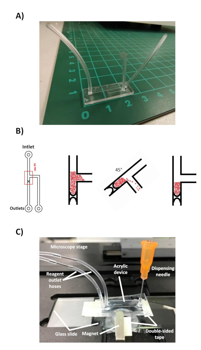
- Keep the device upright to avoid undoing the microparticle trap. See Figure 1B for a summary of the microparticle trap formation process.
3. Immunoassay
- Nanoparticle preparation
- Take 2 µL of the suspension of 100 nm nanoparticles previously conjugated with lysozyme (antigen model). Add it to a 1.5 mL microcentrifuge tube with 100 µL of blocking solution. Incubate overnight at 4 °C.
- Add 150 µL of wash buffer (1x TBS, 0.05% Tween 20).
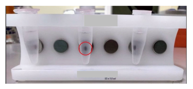
- Place the 1.5 mL microcentrifuge tube in a magnetic separator. Keep for 15 min to allow the separation of the nanoparticles (Figure 2).
NOTE: The minimum volume for the magnetic separator is 200 µL. Avoid using a smaller volume. - Remove the liquid from the tube with a micropipette. Avoid contact with the wall of the tube where the nanoparticle pellet was formed.
- Add 250 µL of fresh wash buffer. Keep the tube under agitation for 15 min.
- Repeat steps 3.1.3.-3.1.5. 2x more, shaking only for 5 min.
- Add the desired concentration of primary anti-lysozyme antibody (see the Table of Materials). Adjust to a final volume of 100 µL in antibody diluent (1x TBS, 1% BSA, 0.05% Tween 20).
- Incubate for 15 min at 37 °C. Keep shaking for an additional 15 min at room temperature.
- Repeat the washing steps 3.1.2.-3.1.6.
- Add 100 µL of antibody diluent. Add the horseradish peroxidase-coupled secondary antibody (HRP-AbII) (see the Table of Materials) at a dilution of 1:500.
- Repeat the washing steps 3.1.2.-3.1.6.
- Keep the nanoparticles in a final volume of 50 µL of antibody diluent.
4. Experimental mounting
- Fill the two 100 µL glass syringes with water, connect a 6.5 cm long hose to each syringe, insert a metal pin into the end of the hose, and place both syringes on the computer-controlled syringe pump.
- Seal all hoses of the acrylic device with heat.
- Cut the inlet hose and keep only a few millimeters. Fill the dispensing needle with wash buffer and insert it into the cut hose. Allow the solution to drip before connecting the needle to the device to prevent air access to the device.
- Cut the outlet hose from the lateral channel. Connect to the syringe pump. Next, do the same procedure for the main channel outlet hose.
NOTE: It is key to perform steps 4.3.-4.4. in this order to avoid unpacking the microparticle trap. If possible, verify the state of the trap during these steps with the help of a magnifying glass. - Place a glass slide on the microscope stage. Attach the magnet to the slide with double-sided tape and place a small piece of tape on each side to fix the chip edges to the glass.
- Set a flow rate of 50 µL/h through the Flow Rate and Units tabs in the syringe pump controller. Select the Withdrawing mode and click on the Start button to activate the flow of the wash buffer.
- Carefully, approach the device toward the slide with the magnet in a horizontal manner so that the area of the chip containing the trap contacts the magnet.
- Stick the edges of the device to the glass with double-sided tape to prevent movement. Avoid obstructing the optical path for microscopy (Figure 1C).
5. Immunodetection
- Keep the wash buffer flowing for 10 min at 50 µL/h to remove excess BSA.
- Remove the remaining wash buffer from the dispensing needle with a micropipette. Add 50 µL of the nanoparticle suspension.
- Flow the suspension of nanoparticles for 7 min at a flow rate of 100 µL/h. Subsequently, change the flow rate to 50 µL/h and flow for another 15 min.
- Change the dispensing needle. Flow the wash buffer for 10 min at 50 µL/h. Prepare the fluorogenic substrate according to the manufacturer's specifications during the washing step.
- Remove the remaining wash buffer from the dispensing needle with a micropipette. Add 100 µL of the fluorogenic substrate (see the Table of Materials). Flow the fluorogenic substrate for 6 min at 50 µL/h.
- Set the flow rate (1 µL/h, 3 µL/h, 5 µL/h, and 10 µL/h) and time (6 min) measurement parameters in the corresponding Flow Rate and Set Timer tabs of the interface that control the syringe pump. Be sure to select Withdrawing mode for each of the measurements to be performed.
- Set an additional Flow Rate tab at 50 µL/h and Set Timer at 3 min for the wash step.
- Turn on the fluorescence of the microscope 15 s before the substrate at 50 µL/h stops. Start the image capture with the software of the microscope camera 10 s before the substrate stops with an exposure time of 1,000 milliseconds. Perform imaging for 6 min at 1 frame/s (FPS).
- Click on the Start button of the desired flow rate parameter immediately after the substrate wash flow rate at 50 µL/h stops. Click on the Start button of the wash flow (50 µL/h) immediately after the selected measurement flow stops.
- Stop the image capture and turn off the fluorescence of the microscope to avoid photobleaching of the substrate.
- Repeat steps 5.8.-5.10. for each measurement flow rate used.
Representative Results

Figure 1: Final device configuration. (A) Acrylic device with the hoses attached to the corresponding inputs and outputs. The scale shows the dimensions of the device in centimeters. (B) Protocol for the formation of the microparticle trap. Microparticles flow through the channel by gravity when the device is placed in a vertical position. Microparticles are concentrated at the 5 µm restriction. Excess microparticles are easily removed by rotating the chip through the side channel. The chip is kept vertical to preserve the trap before immunoassay. (C) Microfluidic device mounted on a glass slide containing the magnet, on the stage of the inverted fluorescence microscope. The dispensing needle through which the reagents are added is observed, as well as the outlet hoses that connect to a syringe pump.

Figure 2: Nanoparticle separation. Using a commercial magnetic separator, 100 nm nanoparticles can be easily concentrated to perform the washing steps during the immunoassay. The pellet formed after 15 min is observed in the red circle.
Divulgations
The authors have nothing to disclose.
Materials
| Carbonyl-iron microparticles | Sigma-Aldrich | 44890 | 7 μm |
| Chloroform | Fermont | 6201 | Health Hazard: Moderate Flammability: None Reactivity: None Contact Hazard: Moderate |
| CMOS camera Moment | Teledyne Photometrics | Sensor Technology: CMOS Quantum Efficiency: 73% Pixel Size: 4.5 µm x 4.5 µm Supported Interfaces: USB 3.2 Gen 2 |
|
| Dr Engrave Software | Roland DGA Corporation | Engraving software to design and create the engraving path on the surface | |
| Extraction hood | Unknown | Unknown | |
| Flexible Plastic Tubing | Tygon | AAD04103 | ID = 0.020, OD = 0.060 |
| Fluorescence microsope | ZEISS | Axio Vert.A1 | |
| High Precision Dispense Needle | Loctite | 98612 | |
| MagJET Separation Rack | thermoscientific | 12 x 1.5 mL | |
| Polymethylmethacrylate – Sheet – PMMA, Acrylic | Goodfellow | ME303018/1 | Thickness: 1.3 mm, Transparency: Clear/Transparent |
| PVCamTest software | Teledyne Photometrics | Version 3.10.107 | Image acquisition software |
| Stereo microscope | Nikon | SMZ 7457 | |
| SuperMag Carboxyl Beads | Ocean NanoTech | KSC0100 | 100 nm |
| Syringe pump | kd Scientific | KDS200 | Can hold up to two syringes |
| Utrasonic bath | Branson | 2800 | |
| VPanel software | Windows OS | Version 1.0.3.0 | Software for controlling the micromilling machine |

