Examining Monosynaptic Connections in Drosophila Using Tetrodotoxin Resistant Sodium Channels
Summary
This article features a method to test the monosynaptic connections between neurons by employing tetrodotoxin and the tetrodotoxin-resistant sodium channel, NaChBac.
Abstract
Here, a new technique termed Tetrotoxin (TTX) Engineered Resistance for Probing Synapses (TERPS) is applied to test for monosynaptic connections between target neurons. The method relies on co-expression of a transgenic activator with the tetrodotoxin-resistant sodium channel, NaChBac, in a specific presynaptic neuron. Connections with putative post-synaptic partners are determined by whole-cell recordings in the presence of TTX, which blocks electrical activity in neurons that do not express NaChBac. This approach can be modified to work with any activator or calcium imaging as a reporter of connections. TERPS adds to the growing set of tools available for determining connectivity within networks. However, TERPS is unique in that it also reliably reports bulk or volume transmission and spillover transmission.
Introduction
A major goal of neuroscience is to map the connections between neurons to understand how information flows through circuits. Numerous approaches and resources have become available to test for functional connectivity between neurons in a range of model systems1,2. To generate the most accurate wiring diagrams using electrophysiology, it is important to resolve whether observed connections between two cells are direct and monosynaptic versus indirect and polysynaptic. A gold standard for making this distinction in mammalian neurons is to measure the latency between single action potentials in the presynaptic cells and the onset of excitatory postsynaptic currents (EPSCs) in the second neuron. Monosynaptic connections should have short latencies of a few milliseconds and low variability3. This approach can be complicated in invertebrate neurons because synapses may occur in their dendrites far from the somatic recording site, causing delays in detection due to long electrotonic distances between postsynaptic conductances and the recording electrode. Such delays can introduce ambiguity regarding poly- versus monosynaptic contributions. Additionally, small synaptic events may decay before reaching the somatic recordings site, and driving stronger presynaptic activity, is likely to recruit polysynaptic events.
Various techniques have been developed to test for monosynaptic connections in invertebrates. One approach uses high divalent cation solutions (Hi-Di) containing excess Mg++ and Ca++. This solution blocks polysynaptic connections by reducing release probability and increasing action potential threshold to favor detection of monosynaptic input4,5. Determining the precise ratio of Mg++ to Ca++ required, however, is not trivial and polysynaptic contributions may persist for even modest stimulation6. An alternative approach called GFP Reconstitution Across Synaptic Partners (GRASP) takes advantage of the proximity between pre- and postsynaptic membranes found at synapses to infer a monosynaptic connection 7. Here, a component of green fluorescent protein (GFP) is expressed in one neuron, and the complementary fragment of the molecule is expressed in a putative postsynaptic partner. The presence of fluorescence indicates that the two neurons are in close enough proximity to permit reconstitution of the GFP molecule and implies the existence of a synapse. GRASP can report synapses falsely, though, if two cells have closely apposed membranes, as in a nerve or fascicle. Variants of GRASP eliminate such false positives by tethering the presynaptic fragment of GFP directly to synaptobrevin, thus allowing reconstitution only at active synapses8. While GRASP and its variants have been instrumental in determining functional connectivity in the Drosophila CNS, some connections may not be made visible by GRASP if the distances between pre- and postsynaptic partners is relatively large. This is particularly pertinent in the assessment of volume transmission associated with neuromodulation9 or GABAergic inhibition10.
Here, a complementary and novel technique is demonstrated for testing direct monosynaptic connections in the Drosophila CNS. This method, called Tetrodotoxin Engineered Resistance for Probing Synapses (TERPS), works by co-expression of the tetrodotoxin (TTX)-resistant sodium channel, NaChBac, and an optogenetic activator in a presynaptic cell while recording from putative postsynaptic partners9. In the presence of TTX, all action potential-mediated activity is suppressed in cells other than those expressing NaChBac. The NaChBac channel selectively restores excitability in the targeted presynaptic cell(s), permitting light-evoked synaptic transmission. This method permits strong activation of the presynaptic neurons, such that connections can be resolved postsynaptically by somatic recording while reducing the probability of recruiting polysynaptic circuits. Importantly, this technique permits the study of volume transmission and reveals the spread of transmitter released from a single neuron throughout a circuit. In addition, TERPS can reveal the chemical nature of the connection through conventional pharmacology. TERPS is suitable for use in any model system that allows the transgenic expression of TTX-insensitive sodium channels.
Protocol
1. Prepare Flies and Identify GAL4 promoter Lines
- Select a GAL4 promoter line and cross it with flies carrying both the UAS-NaChBac transgene and an optogenetic transgene for activation.
NOTE: UAS-CsChrimson11 is selected due to its high conductance and the ease with which it activates NaChBac-mediated action potentials. Both UAS-NaChBac and UAS-CsChrimson are available from the Bloomington Stocks Repository. - Prepare all-trans retinal as a stock solution in ethanol (35 mM) and store the solution in freezer at -20 °C.
- Mix 28 µL of this stock into approximately 5 mL of rehydrated potato flake food and add this mixture to the top of a vial of conventional Drosophila medium.
- Raise adult flies expressing csChrimson on food containing 0.2 mM all-trans-retinal for 1–3 days11.
2. Prepare TTX Stock
- Create a stock solution of 1 mM TTX in distilled or deionized water. Store this solution in a lab refrigerator for several months. Check the institutions policies regarding the storage of TTX as many institutions classify TTX as a hazardous or controlled substance.
3. Prepare Flies for Electrophysiology
- Collect 1–3 day old adult flies that are raised on the food with all-trans-retinal.
NOTE: Refer to Murthy and Turner in 2013 for a detailed description of patch clamping Drosophila neurons12,13. - Put the flies in a glass scintillation vial. Place the vial onto ice for 1 min to immobilize the flies.
- Use coarse forceps to insert a fly into a custom-made recording chamber. The chamber consists of a 3 ½" circle laser cut from 1/16" acrylic. Cut a ¾" hole is into the center where a custom foil is attached with 2-part epoxy. The foil contains a triangular shaped hope that the fly is inserted into.
- Apply wax or UV curable epoxy around the animal to firmly secure it within the recording chamber.
- Illuminate the preparation with a gooseneck optical fiber for visualization under a stereomicroscope. Set the gooseneck fibers for oblique illumination by adjusting them so that they illuminate the fly directly from the side.
- Adjust the light level to lowest possible setting that still allows clear visualization of the fly. This light intensity will vary across individual experiments.
NOTE: This protocol can be adapted by using an in situ brain explant method14. - Retrieve external recording saline. The saline contains in mM: 103 NaCl, 3 KCl, 5 N-tris(hydroxymethyl)methyl-2- aminoethane-sulfonic acid, 8 trehalose, 10 glucose, 26 NaHCO3, 1 NaH2PO4, 1.5 CaCl2, and 4 MgCl2 (adjusted to 270–275 mΩ). The saline is bubbled with 95% O2/5% CO2 (carbogen) and has a pH adjusted to 7.315.
- Place 4-6 drops of extracellular recording saline over the fly preparation. At this point, increase the light level at the dissection microscope for better visibility.
- Sharpen a tungsten wire using electrolysis. To make the wire, prepare a saturated solution of KNO3 and apply approximately 20 V of AC current while dipping the tip of the wire into the solution 20 times for 100 ms each until the tip is sharpened. Use the sharpened tungsten wire to remove the cuticle on the head of the fly and thus exposing the brain.
- Further expose the brain by removing trachea and fat bodies that surround it. If accessing the antennal lobes, use a bent tungsten wire or similar tool to gently tuck antennae underneath the foil recording chamber to increase visibility. Use sharp forceps to gently tear away the glial sheathing covering the area of interest where recordings will be made.
- Transfer the recording chamber to the electrophysiology rig. Immediately start perfusion with external recording saline bubbled with carbogen to preserve the brain. The saline solution reservoir is placed above the microscope stage and is gravity-fed to the preparation. Use a vacuum line and a 2 L Erlenmeyer flask to collect the waste saline.
4. Obtain Whole-cell Recording
- Pull patch pipettes on a commercial pipette puller. Consult the manual for settings to achieve an appropriate pipette with a tip resistance near 7 mΩ.
- Add 4 µL of internal recording solution into a patch pipette. The internal saline consists of 140 potassium aspartate, 10 HEPES, 4 MgATP, 0.5 Na3GTP, 1 EGTA, and 1 KCl in mM.
- Set up patch clamp amplifier for whole-cell recording. Zero the amplifier's offset and set the amplifier to deliver test pulses. Here, an AM systems Patch clamp amplifier 2,400 is used.
- Apply positive pressure through the pipette and approach the cell. Use a micromanipulator to direct the pipette and contact the cell. Release positive pressure. Set the holding potential of the membrane to -60 mV. The pipette should form a gigaohm seal with the cell's membrane as evidenced by a low sub-picoamp holding current.
NOTE: If using GFP guided cell targeting, try to minimize use of epifluorescent light to prevent ChR2 activation and potential synaptic depression. Also wait for a few minutes before applying step 5. - Set the amplifier to deliver test pulses from -50 mV to -60 mV. This setting is standard on patch-clamp amplifiers. Test pulses will reveal a large capacitative transient representing the current necessary to charge the patch pipette. Remove capacitative transients by adjusting the capacity compensation knobs on the AM 2400 or similar amplifier.
- Apply brief pulses of negative pressure to rupture the cell membrane and obtain whole-cell configuration.
NOTE: A whole cell configuration is observed when there is a large increase in the capacitative transients showing the capacitance of the neuron. A small increase in the holding current (baseline) will likely also be observed. Set the amplifier to current clamp mode and adjust the cell's potential to -50 to -60 mV.
5. Test Synaptic Connectivity
- Configure the data acquisition (DAQ) system to trigger an LED driver to generate light pulses. Here, a National Instruments DAQ system is used with Matlab to deliver an analog signal to a LED driver. The LED intensity is adjusted via the analog signal to the driver. The LED is a 620-630 nm wavelength LED.
- Position the high-powered red LED directly underneath the preparation. Adjust the light intensity so that 0.238 mW/mm2 reaches the fly.
- Activate presynaptic neurons while recording from the postsynaptic cell of interest. Achieve cell activation optogenetically, pharmacogentically, or via nerve stimulation. Here, csChrimson and brief pulses of red light are applied.
NOTE: Synaptic connections can be monitored either in current clamp as EPSPs or voltage clamp as EPSCs. A synaptic connection should be visible before applying TTX in response to a 40 ms light pulse. If no connection is observed, it is unlikely that the neurons are synaptically connected either mono- or polysynaptically. The holding potential may be adjusted to emphasize excitatory or inhibitory connections accordingly. - If a connection is observed in normal saline, add TTX to the perfusion system to achieve a final concentration of 1 µM. Use a recirculating pump to capture and reuse the saline to conserve TTX. The peristaltic pump will draw the saline solution from the bath and return it to the saline reservoir.
- Adjust the pump's speed to provide a strong constant vacuum that does not allow that saline level in the bath to rise and fall.
NOTE: The application of TTX should result in the cessation of spiking activity in the neuron that is being monitored in whole-cell. This confirms that enough TTX has been added to the saline. - Apply brief pulses of red light (40 ms for each pulse) again. At this point, any synaptic connection that remains is likely monosynaptic. Add pharmacological antagonists to the recirculating saline to test the chemical nature of the synaptic connections that remain in TTX.
Note: When combining optogenetics and fluorescent targeting of neurons, it may be important not to persistently activate the presynaptic population of neurons while trying to obtain a recording. This can lead to synaptic depression and makes it difficult to see connections. Because csChrimson has a long excitation band that extends broadly into the range of green fluorescent protein, a red fluorophore can be used to visualize neurons and channel2rhodopsin (Ch2R) can be used to excite cell populations. Doing so requires specific genetics and a custom filter cube. To generate suitable flies, make flies that express the red fluorophore (RFP or mCherry) under the LexA or Q system binary expression systems. These will need to be crossed with flies expressing a Gal4 promoter and UAS-Ch2R and UAS-NaChBac effector genes. The optimal filter cube for epifluorescence will have a narrow excitation range to excite the red fluorophore, but not excite the Ch2R. Additionally, the filter set should include a long pass emission filter to collect as much emitted light. We used a custom filter set from Chroma that included part numbers et580/25x and t600lpxr (from the 49306 set), but with an et610lp barrier/emission filter.
Representative Results
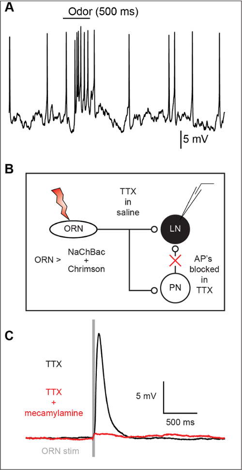
TERPS is used to distinguish between mono- and polysynaptic contributions in synaptic connections between neurons. While weak stimulation of a cell may be used to test direct connections, driving greater presynaptic activity often recruits polysynaptic connections (Figure 1A). TERPS works by co-expressing the TTX-insensitive sodium channel NaChBac and an optogenetic activator, and testing connections in the presence of TTX to eliminate polysynaptic connections (Figure 1B). TTX effectively blocks action potentials in the Drosophila brain, making it a suitable preparation for TERPS (Figure 2A). Transgenic expression of the NaChBac sodium channel rescues excitability in neurons and results in a large plateau potential (Figure 2B).
Local interneurons (LN) in the antennal lobe show robust responses to olfactory stimulation (Figure 3A). However, because individual EPSPs are not easily resolved at the LN soma, it remains unclear if such responses arise directly from olfactory receptor neuron input (ORN) or indirectly input via projection neurons (PN) (Figure 3B). By using TERPS, ORNs shown here, do indeed make direct synaptic connections with LNs (Figure 3C).
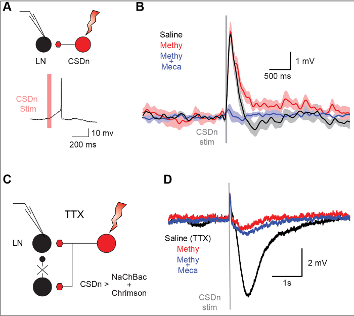
A unique feature of TERPS is its ability to specifically resolve extra-synaptic volume transmission that may occur with either GABA or neuromodulators such as serotonin. Stimulation of a serotonergic neuron (the CSDn) in the Drosophila antennal lobe results in a mixture of excitation and inhibition in a local interneuron (Figure 4A and Figure 4B). The excitation is meditated by the excitatory transmitter acetylcholine and the inhibition is caused by serotonin (Figure 4C and Figure 4D). TERPS can be used to determine if the connections are monosynaptic by eliminating polysynaptic connections (Figure 4E). In TTX, a strong inhibition is still observed in the LN, suggesting it arrives from directly from the activation of a specific serotonergic neuron (Figure 4F and Figure 4G). This connection is blocked by the serotonin antagonist methysergide. The excitatory synapse mediated by acetylcholine was eliminated in TTX, suggesting it arose from polysynaptic sources.

Figure 1: TERPS eliminates polysynaptic connections and isolates monosynaptic inputs. (A). A schematic diagram showing that increasing presynaptic activity can recruit polysynaptic connections. (B). A diagram illustrating how TERPS can remove polysynaptic contributions by using TTX to eliminate spiking activity in neurons. Excitability is exclusively restored in one population of neurons that can then be excited optogenetically. Please click here to view a larger version of this figure.

Figure 2: NaChBac restores neuronal excitability in Drosophila neurons. (A) The application of 1 µm TTX abolishes spiking in Drosophila neurons. Both spontaneous activity and odor-evoked inhibition are eliminated in TTX. (B) NaChBac and csChrimson are expressed in the CSDn and the brain is exposed to TTX. csChrimson stimulation depolarizes the CSDn and after a threshold is crossed, there is large non-linear increased in the CSDn membrane voltage. This rapid depolarization constitutes a plateau-like potential (inset). Each color across simulation intensities corresponds to the same color in the voltage inset. This figure has been modified from Zhang and Gaudry 20169. Please click here to view a larger version of this figure.

Figure 3: TERPS reveals monosynaptic connections between ORNs and LNs. (A) A sample recording showing an odor response in an LN. This response may be mono- or polysynaptic in nature. (B) TERPS can be tested at the ORN to LN synapse. TTX is used to block action potentials and excitability in all cells in the preparation. Excitability is exclusively restored in the ORNs via the NaChBac sodium channel. The ORNs can then be excited with channelrhodopsin to elicit synaptic release. Synaptic events are measured post-synaptically using whole-cell recordings. (C) TERPS reveals that the ORN to LN synapse is monosynaptic. TERPS can also be combined with pharmacology to show that the synapse is cholinergic and blocked by the nicotinic receptor antagonist mecamylamine (200 µM). The vertical gray bar indicates the timing of ORN stimulation. This figure has been modified from Zhang and Gaudry in 20169. Please click here to view a larger version of this figure.

Figure 4: TERPS is sensitive to bulk or volume transmission release from modulatory neurons. (A) The stimulation of the CSDn results in an action potential from a recorded LN in the Drosophila antennal lobe. (B) The LN is hyperpolarized so that CSDn stimulation results in a subthreshold activity. The LN response consists of a fast depolarization followed by a slower hyperpolarization. The gray bar denotes the time of CSDn stimulation. Methysergide (50 µM), a broad 5-HT receptor antagonist, blocks the slow hyperpolarization but has no effect on the depolarization. Mecamylamine blocks the fast depolarization suggesting that it is cholinergic in nature. (C) TERPS can be used to resolve which chemical components of the CSDn to LN synapse are mono- versus polysynaptic. (D) The CSDn is stimulated in the presence of TTX and postsynaptic LN responses are measured in whole-cell recordings. The hyperpolarization persists in TTX suggesting it is monosynaptic in nature. This hyperpolarization is also blocked by methysergide, consistent with serotonergic transmission. The brief depolarization that remains in TTX is likely mediated by electrical gap coupling, as it not blocked by nicotinic antagonists. This figure has been modified from Zhang and Gaudry in 20169. Please click here to view a larger version of this figure.
Supplementary File 1: Drosophila Foil. Please click here to download this file.
Supplementary File 2: Physiology Chamber. Please click here to download this file.
Discussion
TERPS analysis compliments current techniques used for circuit cracking by enabling the detection of synapses between identified neurons. Specifically, the approach reveals monosynaptic connections by broadly silencing action potentials with TTX while restoring excitability in a select population of neurons with the TTX-insensitive sodium channel NaChBac. Synaptic release is elicited by optogenetic stimulation while postsynaptic events are monitored with whole-cell recordings. TERPS distinguishes itself from other approaches such as GRASP by being sensitive to bulk or volume transmission elicited at longer distances and the ability to perform pharmacological manipulations. The technique is relatively straightforward to employ and requires only one recording electrode and a light source for optogenetic stimulation, which is more feasible than obtaining dual whole-cell recordings on both pre- and post-synaptic parts of the synapse. A basic requirement of TERPS is that action potentials are easily blocked by TTX in the system being studied. But, because TTX acts on neurons across a wide range of taxonomic phyla, it is likely that TERPS could be applied broadly to both mammalian and invertebrate model systems.
TERPS can be easily modified in a number of manners to facilitate either the stimulation of the putative presynaptic population or for recording from postsynaptic targets. Here, an optogenetic tool was used to stimulate the target populations, though activation by a variety of mechanisms is possible. Nerve stimulation of sensory axons, for example, is routine in studies of transmission from the antennal nerve in Drosophila16 and is suitable in other sensory systems, including peripheral nerves containing mechanosensory neurons. A similar approach to testing functional connectivity in Drosophila expressed GCaMP calcium indicators in one population of neurons while driving activity in another population via the ionotropic purinoceptor P2X2 and ATP application2. This approach should be compatible with TERPS by simply co-expressing the NaChBac transgene with the P2X2 receptor in one neuron and GCaMP in another population for a completely patch-free method. However, caution should be taken with approaches causing slower depolarizations, such as the muscarinic-based designer receptors exclusively activated by a designer drug (DREADDs)17,18. Slow depolarization could lead to NaChBac inactivation prior to action potential generation.
Similar methods to TERPS have been employed in mammalian systems. Such techniques combine TTX and channelrhodopsin to map out connectivity between neurons. Channelrhodopsin activation alone generally does not depolarize neurons sufficiently, and thus the potassium channel blocker 4-AP is be applied with TTX to elicit release19,20,21. While this works at many synapses, it may not work at some metabotropic synapses if the downstream target of the receptor is a 4-AP sensitive potassium channel. For instance, serotonergic modulation of olfactory response in moths relies directly on modulation of such a 4-AP sensitive K+ current (IA)22. This approach also did not work at the CSDn to LN synapse (data not shown). Thus, TERPS has a benefit of requiring less pharmacological intervention compared to other commonly used methods and may work at wider range or synapses.
There are a few limitations to TERPS that should be considered. First, there may be potential side effects from chronic expression of the NaChBac channel throughout development. Proper circuit assembly and function can be confirmed by comparing the activity of neurons between control flies lacking the NaChBac construct and flies with the construct in the absence of TTX. To avoid any such problems, expression of the NaChBac transgene could be controlled temporally via expression of the temperature-sensitive GAL4 repressor, GAL8023. Additionally, if a synapse is only observed in a state-dependent manner, it is conceivable that suppressing network activity with TTX might conceal such a connection. It is important to emphasize that a positive connection observed with TERPS likely represents a true mono-synaptic connection, while a negative result is not proof of the absence of a connection.
While TERPS can reveal the ability of transmitter release from one neuron to affect postsynaptic partner clearly, the slow channel dynamics of NaChBac and the plateau potentials associated with its activation render it less applicable to the study of classical neurotransmission. One approach to altering TERPS for faster, more natural transmitter release will be to express a modified TTX-insensitive version of the endogenous Drosophila sodium channel para as opposed to NaChBac. This alteration would result in naturalistic spiking activity in the presynaptic cell and permit more conventional analysis of synaptic transmission in the absence of network activity. This would have great potential for studies of rate-coding synapses, at which eliciting a broad range of presynaptic activity without recruiting polysynaptic networks would be desirable.
Offenlegungen
The authors have nothing to disclose.
Acknowledgements
We would like to thank Joshua Singer, Jonathan Schenk, as well as thoughtful reviewers for comments on the manuscript. We would also like to thank Ben White and Harold Zakon for discussions on the technique. Jonathan Schenk provided the data for Figure 3A. This work was supported by a Whitehall Foundation Grant and an NIH R21 to QG.
Materials
| UAS-csChrimson | Bloomington Drosophila Stock Center | 55135 | Used as a neural activator |
| UAS-NaChBac | Bloomington Drosophila Stock Center | 9466 | Resotores excitibility in cells in TTX |
| Tetrodotoxin | Tochris | 1078 | Special permission may be needed to purchase TTX as it is a controlled substance |
| all trans-Retinal | Sigma-Aldrichall trans-Retinal | R2500 | Require co-factor for channelrhodopsin |
| Weldable 321 Stainless Steel Sheet, 0.002" Thick, 10" Wide | McMaster Carr | 3254K7 | Used to make custom fly holder. Custom foil can be laser cut at pololu.com from the provided PDF file |
| Dissection Microscope | Zeiss | Stemi 2000-C | Used for dissection of preparation |
| Waxer | Almore | Eectra Waxer 66000 | Used during dissection to secure fly in foil |
| Paraffin Wax | Joann | 4917217 | Used with waxer |
| Number 5 forceps | Fine Science Tools | #5CO | Used for dissection and desheathing |
| Dissection Scissors | Fine Science Tools | 15001-08 | Used to remove parts of the cuticle during dissection |
| Tungsten wire | A-M Systems | 797500 | Use with electrolysis to make sharpened needles for dissection |
| Reciculating Peristaltic Pump | Simply Pumps (Amazon) | PM200S | for recirculating TTX |
| Speed Controller for peristaltic pump | Zitrades (Amazon) | N/A | PWM Dimming Controller For LED Lights or Ribbon, 12 Volt 8 Amp,Adjustable Brightness Light Switch Dimmer Controller DC12V 8A 96W for Led Strip Light B |
| Versa-Mount Precision Compressed Air Regulator | McMaster Carr | 1804T1 | For applying positive pressure during patching |
| Glass capilaries | World Precision Instruments | TW150F-3 | For patch pipettes |
| Multipurpose Gauge | McMaster Carr | 3846K431 | Gauge for pressure regulator |
| Electrophysiology Camera | Dage MTI | IR-1000 | Any camera that works in the IR range (850 nm) will work. You do not want to use red illumination as this can activate csChrimson |
| IR LED | Thorlabs | M850F2 | For oblique illumination |
| fiber optic for IR LED | Thorlabs | M89L01 | Couples to IR LED |
| Objective lens | Olympus 40X | LUMPlanFLN | This can be used on most microscipes and works well for visualizing fly neurons. |
| Amber LED | Thorlabs | M590L3d | For visualizing RFP and mCherry |
| Blue LED | Thorlabs | M470L3d | For visualizing GFP |
| GFP filter set | Chroma | 49011 | For visualizing GFP or stimulating channel2rhodopsin |
| Custom mCherry Filter Set | Chroma | et580/25x and t600lpxr (from the 49306 set) but with an et610lp barrier/emission optic | Use only if you wish to patch identified neurons with channel2rhodopsin |
| Dichroic to combine Amber and blue LED | Thorlabs | DMLP550R | Use only patch under mCherry and excite channel2rhodopsin with blue light. |
| Red LED | LEDSupply | Cree XPE 620 – 630 nm | Used to drive csChrimson |
| LED Driver | LEDSupply | 3021-D-E-1000 | Used to drive LEDs for optogenetic stimulation |
| Manipulator | Sutter Instruments | MP-225 | Used to position pipette during recordings |
| Patchclamp Amplifier | A-M Systems | Model 2400 | An equivalent amplifier is suitable |
| Bessel Filter | Warner Instruments | LPF 202A | Auxillary filter used to filter current trace to oscilloscope during patching. |
| Data Acquisition System | National Instruments | NI PCIe-6321 781044-01 | Used to record data from amplifier to computer |
| Connector Block – BNC Terminal BNC-2090A | National Instruments | 779556-01 | Used to connect amplifier to DAQ card. |
| Steel Foil | McMaster Carr | 3254K7 | Steel foil for custom recording chamber |
| Magnets | K&J Magnetics | D42 | To secure recording chamber to ring stand |
| 1/16" Cell Cast Acrylic Clear | Pololu | Used to make custom recording chamber. Acrylic can be laser cut at pololu.com from the provided PDF file |
Referenzen
- Petreanu, L., Huber, D., Sobczyk, A., Svoboda, K. Channelrhodopsin-2-assisted circuit mapping of long-range callosal projections. Nat Neurosci. 10, 663-668 (2007).
- Yao, Z., Macara, A. M., Lelito, K. R., Minosyan, T. Y., Shafer, O. T. Analysis of functional neuronal connectivity in the Drosophila brain. J Neurophysiol. , 684-696 (2012).
- Doyle, M. W., Andresen, M. C. Reliability of monosynaptic sensory transmission in brain stem neurons in vitro. J Neurophysiol. 85, 2213-2223 (2001).
- Nicholls, J. G., Purves, D. Monosynaptic chemical and electrical connexions between sensory and motor cells in the central nervous system of the leech. J Physiol-London. 209, 647-667 (1970).
- Byrne, J. H., Castellucci, V. F., Kandel, E. R. Contribution of individual mechanoreceptor sensory neurons to defensive gill-withdrawal reflex in Aplysia. J Neurophysiol. 41, 418-431 (1978).
- Liao, X., Walters, E. T. The use of elevated divalent cation solutions to isolate monosynaptic components of sensorimotor connections in Aplysia. J Neurosci Meth. 120, 45-54 (2002).
- Feinberg, E. H., et al. GFP Reconstitution Across Synaptic Partners (GRASP) Defines Cell Contacts and Synapses in Living Nervous Systems. Neuron. 57, 353-363 (2008).
- Macpherson, L. J., et al. Dynamic labelling of neural connections in multiple colours by trans-synaptic fluorescence complementation. Nat Commun. 6, 10024-10029 (2015).
- Zhang, X., Gaudry, Q. Functional integration of a serotonergic neuron in the Drosophila antennal lobe. Elife. 5, (2016).
- Wilson, R. I. Early olfactory processing in Drosophila: mechanisms and principles. Annu Rev Neurosci. 36, 217-241 (2013).
- Klapoetke, N. C., et al. Independent optical excitation of distinct neural populations. Nat Methods. 11, 338-346 (2014).
- Murthy, M., Turner, G. C. Whole-cell in vivo patch-clamp recordings in the Drosophila brain. Cold Spring Harb Protoc. , 140-148 (2013).
- Murthy, M., Turner, G. C. Dissection of the head cuticle and sheath of living flies for whole-cell patch-clamp recordings in the brain. Cold Spring Harb Protoc. 2013, 134-139 (2013).
- Gu, H., O’Dowd, D. K. Whole Cell Recordings from Brain of Adult Drosophila. J Vis Exp. , (2007).
- Wilson, R. I., Turner, G. C., Laurent, G. Transformation of olfactory representations in the Drosophila antennal lobe. Science. 303, 366-370 (2004).
- Kazama, H., Wilson, R. I. Homeostatic Matching and Nonlinear Amplification at Identified Central Synapses. Neuron. 58, 401-413 (2008).
- Becnel, J., et al. DREADDs in Drosophila: a pharmacogenetic approach for controlling behavior, neuronal signaling, and physiology in the fly. Cell Reports. 4, 1049-1059 (2013).
- Armbruster, B. N., Li, X., Pausch, M. H., Herlitze, S., Roth, B. L. Evolving the lock to fit the key to create a family of G protein-coupled receptors potently activated by an inert ligand. P Natl Acad Sci USA. 104, 5163-5168 (2007).
- Sun, Q. Q., Wang, X., Yang, W. Laserspritzer: A Simple Method for Optogenetic Investigation with Subcellular Resolutions. PLoS One. 9, e101600-e101608 (2014).
- Holloway, B. B., et al. Monosynaptic Glutamatergic Activation of Locus Coeruleus and Other Lower Brainstem Noradrenergic Neurons by the C1 Cells in Mice. J Neurosci. 33, 18792-18805 (2013).
- Petreanu, L., Mao, T., Sternson, S. M., Svoboda, K. The subcellular organization of neocortical excitatory connections. Nature. 457, 1142-1145 (2009).
- Kloppenburg, P., Ferns, D., Mercer, A. R. Serotonin enhances central olfactory neuron responses to female sex pheromone in the male sphinx moth manduca sexta. J Neurosci. 19, 8172-8181 (1999).
- Suster, M. L., Seugnet, L., Bate, M., Sokolowski, M. B. Refining GAL4-driven transgene expression in Drosophila with a GAL80 enhancer-trap. Genesis. 39, 240-245 (2004).

