Establishment of Larval Zebrafish as an Animal Model to Investigate Trypanosoma cruzi Motility In Vivo
Summary
In this protocol, fluorescently labeled T. cruzi were injected into transparent zebrafish larvae, and parasite motility was observed in vivo using light sheet fluorescence microscopy.
Abstract
Chagas disease is a parasitic infection caused by Trypanosoma cruzi, whose motility is not only important for localization, but also for cellular binding and invasion. Current animal models for the study of T. cruzi allow limited observation of parasites in vivo, representing a challenge for understanding parasite behavior during the initial stages of infection in humans. This protozoan has a flagellar stage in both vector and mammalian hosts, but there are no studies describing its motility in vivo.The objective of this project was to establish a live vertebrate zebrafish model to evaluate T. cruzi motility in the vascular system. Transparent zebrafish larvae were injected with fluorescently labeled trypomastigotes and observed using light sheet fluorescence microscopy (LSFM), a noninvasive method to visualize live organisms with high optical resolution. The parasites could be visualized for extended periods of time due to this technique's relatively low risk of photodamage compared to confocal or epifluorescence microscopy. T. cruzi parasites were observed traveling in the circulatory system of live zebrafish in different-sized blood vessels and the yolk. They could also be seen attached to the yolk sac wall and to the atrioventricular valve despite the strong forces associated with heart contractions. LSFM of T. cruzi-inoculated zebrafish larvae is a valuable method that can be used to visualize circulating parasites and evaluate their tropism, migration patterns, and motility in the dynamic environment of the cardiovascular system of a live animal.
Introduction
Chagas disease is caused by the protozoan parasite T. cruzi. Approximately 6 to 7 million people worldwide are infected with T. cruzi. The disease is transmitted mainly in Latin America, but has been reported in the United States, Canada, and many European as well as some Western Pacific countries, mainly due to migration of infected individuals1. Chagas is largely vector-borne and transmitted to humans by contact with the feces of hematophagic insects in the Triatominae subfamily, commonly known as "kissing bugs". However, T. cruzi can also be transmitted via blood transfusions, vertical transference from mother to child, or ingestion of food contaminated with parasites2. The acute phase the infection is mainly asymptomatic or constitutively symptomatic and lasts from 6 to 8 weeks, after which engagement of the immune system controls parasite load, but does not completely eliminate the infection3. Most individuals then enter a chronic asymptomatic phase; however, nearly 30% of patients develop a symptomatic chronic phase, in which the cardiac system and less frequently the digestive and nervous systems are compromised4. This scenario presents a challenge for disease treatment and control since there are no vaccines available, and there are only two effective drugs for Chagas: benznidazole and nifurtimox. Both treatments require prolonged administration and may have severe side effects2.
Increased understanding of the behavior of T. cruzi behavior in vivo is key to determining parasitic migration, cellular attachment, and invasion within the host; a lack of in vivo models limits the development of novel therapeutic approaches. In vitro studies of T. cruzi infection have shown that motility of trypomastigotes is important for binding to host cell membranes and subsequent cellular invasion5. Energy depletion in trypomastigotes in co-culture with a susceptible cell line has been shown to reduce cellular invasion6. Interestingly, in trypanosomatids, flagellar movement has also been characterized as an evasion mechanism against parasite-specific antibodies7.
Flagellar motility has been extensively studied in vitro in Trypanosoma brucei, a closely related parasite that causes African Trypanosomiasis8. In vitro studies of the motility of these trypanosomes showed that simulation of the conditions of blood or body fluids, including viscosity and the presence of obstacles representative of blood cells, is important for parasite forward movement9. As of yet it has not been possible to visualize the movement of parasites in the bloodstream in vivo.
Zebrafish larvae are a powerful model to study host-pathogen interactions in vivo. They are small, inexpensive, and relatively easy to raise when compared with other established vertebrate models for Chagas disease. Zebrafish have innate and adaptive immune systems similar to humans, but their adaptive immune system begins to develop at 4 days post fertilization (dpf) and is not mature for another several weeks10. During early development, when only macrophages are present, there is a large window for studying parasite behavior without immediate immune interference10. However, the greatest advantage of utilizing zebrafish larvae as a vertebrate model for studying pathogen behavior lies in their optical transparency, making them amenable for microscopic screening and imaging11. Additionally, there are multiple tools to manipulate fish genetics. For example, the Casper strain is a mutant line of zebrafish with no pigmentation, making the animal completely transparent and useful for visualization of individual organs and for real-time tracking of injected cells12.
A key limitation of longitudinal observation of swiftly moving parasites in live zebrafish using confocal or epifluorescence microscopy lies in the impossibility of imaging at high acquisition speeds and large penetration depths with good image quality and low risk of photodamage. Light sheet fluorescence micsroscopy (LSFM) is an emerging imaging technique that overcomes these limitations to permit these observations. By using one objective to detect fluorescence and a second orthogonal illumination objective that only illuminates the focal plane of the detection objective, it is possible to obtain high resolution optical sections as in a confocal microscope, but with significantly reduced photodamage, even with respect to epifluorescence microscopy13. The LSFM technique used here is called Single Plane Illumination Microscopy (SPIM), in which a thin sheet of light illuminates a single plane within the zebrafish larvae.
The objective of this methodology is to establish larval zebrafish as a viable non-infection model for understanding T. cruzi motility and related behavior in vivo. To accomplish this, we injected transparent zebrafish larvae with fluorescently labeled trypomastigotes, the cellular form responsible for infection of humans, and identified the movement of T. cruzi in the cardiovascular circulation of zebrafish using LSFM.
Protocol
The following protocols were approved by the Institutional Animal Care and Use Committee of Los Andes University (CICUAL). Biosafety level 2 (BSL-2) guidelines should be strictly followed to prevent contamination with the pathogen T. cruzi.
Note: Animal care and maintenance: Casper zebrafish, a genetically modified strain of zebrafish (Danio rerio) is used in this protocol due to their valuable optical transparency in all developmental stages. Fish are manipulated using optimal care conditions for the species14, in a 14 h light-10 h dark cycle, at 28 ± 1 °C, in a pH (7.0-7.4) controlled multi-tank recirculating water system. Animals are fed twice a day with live brine shrimp (Artemia salina) and enriched with rearing food. All protocols were approved by CICUAL at Universidad de los Andes (C.FUA_14-017).
1. Preparation of Egg Water
- Prepare 0.6 g/L aquarium salt in reverse osmosis (RO) or deionized (DI) water and add 0.01 mg/L methylene blue to the solution.
Note: The measured conductivity should be of 400-500 µS/cm and pH level at 7.2-7.9. To lower the pH, aerate the egg water for a few hours.
2. Preparation of Tricaine Stock Solution
- Prepare 0.4% tricaine stock solution by dissolving 400 mg tricaine (MS-222) in 97.9 mL distilled water (ddH2O). After the powder has completely dissolved, adjust the pH to 7.0 using 2.1 mL 1M Tris (pH 9.0) and refrigerate the solution.
3. Preparation of 1.0% Low Melting Point Agarose
- Dissolve the low melting point agarose powder in egg water to a final concentration of 1.0%. Heat the mixture in a microwave or on a hot plate with continuous stirring until the agarose solution appears homogeneous.
- Store the aliquots at 4 °C for no longer than a week.
4. Mating Assay and Embryo Collection
- Three days prior to injections set up matings using healthy pairs of male and female fish in breeding tanks. Enrichment with glass marbles and artificial plants enhances spawning. Matings should be set up during the afternoon and left overnight.
- The next morning, collect the spawned eggs by draining the tank through a strainer and washing the eggs with the egg water. Remove all the eggs by inverting the strainer and pouring egg water through the strainer into a Petri dish. To keep the embryos healthy, clean their water by removing any debris and dead embryos with a transfer plastic pipette.
- Transfer the embryos to an incubator at a stable temperature of 28.5 °C to allow development of the embryos according to the zebrafish staging standards15. Examine the embryos two times a day, and discard inviable eggs to keep the clutch healthy.
5. Embryo Dechorionation
Note: This procedure is required if the embryos have not hatched at the time of injection. In this procedure, "larvae" are animals out of their chorion from 48 h post fertilization (hpf) onward.
- On the day of the experiment, place the Petri dish containing the healthy embryos under a dissecting stereoscope.
- Grab opposing ends of the chorion with two sharp forceps (Dumont #5), and gently tear and pull open the chorion. About 5-6 larvae are injected at a time, and typically 3-4 are imaged.
- Remove the chorions from the water using a transfer pipette.
6. Preparing Injection Material
- Prepare 1.0 mm needles using thin wall glass capillaries and a micropipette puller device with the following settings: 2 light weights + 1 heavy weight, 2 mm top pull + 5 mm bottom pull, 75.9 °C (Step 1) + 78.2 °C (Step 2). Store needles in a Petri dish on a strip of modeling clay. The ideal needle should have a narrow tip, as T. cruzi measure approximately 20 x 1-3 µm and will pass through the needle easily. The length of the tip can vary: a longer tip is useful to reach deeper structures, but a shorter tip will be more rigid making injection easier for the user.
Note: The needle tip size depends on the temperature used: a higher temperature for Step 2 results in a longer and finer tip. - Prepare a 1.5% agarose solution in ddH2O and pour it into a 10 cm Petri dish. Cover a depth of approximately 1.0 cm.
- Place a prefabricated microinjection mold over the agarose and allow it to solidify.
- Lift the prefabricated microinjection mold out and add egg water for storage at 4 °C. This prevents the agarose from drying out.
- Add egg water to the tricaine stock solution to a final concentration of 150 mg/L. Store at 4 °C.
7. Cell Culture for Parasite Growth
- Parasites are maintained in a human astrocytoma cell line (CRL-1718) using the method described by Vargas-Zambrano et al.16
- Start the culture of human cells in a T25 culture flasks (surface area = 25 cm2) at a density of 2 x 10⁵ or 4 x 10⁵ cells in 4 mL of RPMI-1640 medium with 10% of fetal calf serum (FCS), supplemented with 2 mM L-glutamine, 4.5 g/L glucose, 10 mM HEPES, 1 mM pyruvate, and 1% penicillin-streptomycin (referred as "complete media"). Incubate the astrocytoma cultures at 37 °C in a 5% CO2 environment.
- Once a monolayer is confluent, detach the cells using 2 mL of 0.25% of trypsin-EDTA by incubating them for 3 min at 37 °C. Visually check cells for detachment, and block the trypsin solution using 3 mL of RPMI-1640 with 10% FCS in a 15 mL tube.
- Centrifuge the cell suspension for 5 min at 1,350 x g.
- Wash the resulting cellular pellet in DMEM medium in a 15-mL tube and centrifuge for 5 min at 1,350 x g at 22 °C.
- Count the cells manually in a Neubauer chamber and check for viability in a light microscope at 40X magnification.
- Gently re-suspended the cellular pellet in 4 mL of complete media and use it to create new cell cultures using 2 x 10⁵ cells per T25 culture flask.
8. T. cruzi Culture and Labeling
- Parasites are a strain originally obtained from an infected human and characterized as T. cruzi DA strain (MHOM/CO/01/DA), genotype Discrete Typing Unit (DTU) TcI16. After 3 or 4 days of culturing, collect the moving parasites from the human astrocytoma cell culture supernatant and centrifuge for 5 min at 1,350 x g. Discard the medium.
Note: Parasites in complete medium can be used for re-culture in a 1:1 ratio with astrocytoma cells in T25 culture flasks. - Gently re-suspend the pellet in 10 mL sterile 1x Phosphate Buffer Saline (PBS) with 0.1% FCS and centrifuge for 5 min at 1,350 x g.
- Discard the supernatant and re-suspend in 1 mL of PBS. Take 10 µL for counting the free-swimming parasites in a Neubauer chamber.
- Take the rest of the re-suspended parasites (990 µL) and add 1 µL of Carboxyfluorescein Diacetate Succinimidyl Ester (CFSE), final concentration 5.0 µM. Incubate for 10 min at room temperature.
Note: CFSE stock at 5 mM should be aliquoted and maintained at -20 °C. - Pellet the parasites (1,350 x g, 5 min). Reconstitute the parasite pellet by flicking the tube and wash in 10 mL of 1x PBS-0.1% FCS followed by a subsequent spin (1,350 x g, 5 min).
- Discard the supernatant and gently re-suspend the labeled parasite pellet in 1X PBS with the appropriate volume to obtain a final concentration of about 10-20 parasites/nL for injection.
- Use a small volume (∼10 µL, approximately 1 x 10⁴ -2 x 10⁴ parasites) of trypomastigotes to assess viability and labeling. An inverted fluorescence microscope can be used for direct visualization of the parasites. In the transmitted light mode, check that the parasites move. In the fluorescence mode, use the Fluorescein Isothiocyanate (FITC) filter to assess parasite labeling.
9. Injecting Zebrafish Larvae
- Parasite loading
- Take 10 µL of parasites from the resuspension (100 µL) and load them into the sealed glass needles using a 10 µL microloader pipette tip.
- Insert the glass needle into the needle holder of the micromanipulator, use a magnetic stand, and place next to the stereoscope.
- Using fine forceps under the stereoscope, cut the needle creating a blunt open end and measure the drop size to obtain an injection volume of 1 nL (this is equivalent to a bead diameter of 0.12-0.13 mm when measured in mineral oil17). A longer tip is preferred so that the needle can reach structures deep inside the fish without damaging the tissue. In this protocol, a blunt needle is used, but a beveled needle can be used as well.
- Mounting
- Anesthetize larvae of 48 hpf, with 150 mg/L tricaine. Check that they do not respond to touch, but ensure that the heart is beating.
- Place the larvae in the microinjection agarose mold in a lateral position.
- Place the needle holder next to the stereoscope and adjust the micromanipulator so that the tip of the needle is in the center of the field of view. Move the Petri dish prepared in steps 6.2- to 6.4 so that the larval yolk is close to the needle tip. Check the condition of the larva to ensure that the heart continues beating.
- Set the microinjector values as follows: injection pressure 9.6 pounds per inch (psi); hold pressure: 20 psi; 100 ms range of gating; period value of 1.9 (corresponds to 10.9 ms). The volume of the injection should be between 1-3 nL, with approximately 10-20 parasites/nL.
10. Injection of the Parasite
- Inject the fish in the superior-anterior portion of the yolk at the duct of Cuvier. This step should be practiced to ensure animal survival after injection. It takes approximately 10 min to inject 5-6 larvae successfully.
- As a control, inject 1-2 larvae with the same vehicle volume (1x PBS).
- Transfer the larva to fresh egg water immediately.
11. LSFM Mounting of Injected Larvae
- Transfer the larva injected with parasites to an empty Petri dish and carefully remove the surrounding water with a plastic pipette and absorbent paper. Immediately add 100 µL of preheated 1.0% low melting point agarose (refer to step 3 for agarose preparation) to cover the larva. Ensure the agarose is not over 40 °C.
- Insert a straight wire inside a 1.0 mm glass capillary and use it as a plunger to suck up the larva in a vertical position. Make sure to leave a small amount of agarose above the larva. Wait until the agarose solidifies before exposing the larva (this will require a few minutes). If necessary, push out excess agarose below the larva and cut it off.
- Place the capillary in the microscope sample holder and push out the end containing the larva until it hangs free from the capillary. The specimen chamber should be filled with tricaine solution (150 mg/L) and the temperature set at 28 °C.
- Position the larva in front of the pupil of the detection objective using an XYZ-micromanipulator system. For rotations about the vertical axis, use a rotation stage. Positioning of the larva should be done in transmitted light mode to clearly identify larval structures.
12. LSFM Imaging of Injected Parasites
- Change to fluorescence mode and adjust the illumination intensity as well as the exposure time to decrease photodamage and optimize time resolution. For these particular experiments, we used a power of 2.8-3.0 mW for the sample and exposed each frame for 200 ms using a camera with 6.45 µm pixels and ~70% quantum efficiency at the detection wavelengths. These settings should result in adequately exposed images at about 5 frames/s. Make sure to use an objective with the highest possible numerical aperture.
- Start a video acquisition of the region of interest (ROI). In our case, parasites are observed attached to valves and freely moving around the heart area. It is recommended to take a video of a single plane over time, or to use the micromanipulator or piezo-galvo system to focus different planes while the parasite moves.
Note: This procedure is useful for short acquisition times of up to 2 h. For long term acquisition, the larvae should be mounted using alternative methods20.
13. Image Processing and Analysis of Acquired Data
Note: Image processing was performed on a personal computer with a 2.90 GHz processor, 8.00 GB of memory, and a videocard with 1.00 GB of memory.
- Open the acquired dataset in the image processing software of choice. The open software FiJi is recommended for LSFM data processing and analysis21.
- In the image analysis software, adjust brightness and contrast levels to enhance the images.
Note: No fixed parameters are used in this case, but selecting the "Auto" option can be useful to obtain an initial enhancement. - Select the ROI.
Note: For tracking individual parasites, FiJi has both manual and automatic tracking plugins available.
14. Recovering Imaged Larvae
- Remove the larva carefully from the agarose using fine forceps and a hair loop tool. Transfer the fish back to fresh egg water and check for recovery after 15 min. Return the larva to incubator at 28 ± 0.5 °C.
- Alternatively, proceed to euthanize the imaged larvae with an overdose of tricaine. Then, introduce the larvae in a solution of sodium hypochlorite (6.15% NaClO) for 5 min to kill the parasite. Dispose according to the institution's standard protocols.
Representative Results
Optimal Conditions for Injection:
Groups of zebrafish larvae were injected at 24, 48, 72, 96, and 120 hpf, at different anatomical sites, and their survival was examined every day for 5 days. After 5 days post injection, embryos injected at 24 hpf had 6.25% (2/32) survival, whereas 95% (38/40) of larvae injected at 48 hpf survived. As a control, larvae were injected with 1x PBS as a vehicle. There were no differences in survival between vehicle-injected and parasite-injected larvae, indicating no parasitic-dependent effects on survival rate of the fish (p = 0.08). Larvae injected between 72-120 hpf had comparable survival rates to 48 hpf-injected larvae at constant injection volume. For all procedures presented here, 48 hpf larvae were used due to their ease of manipulation and having developed organs and easily penetrable skin without evident damage after injection.
Larvae injected at 48 hpf were injected in the pericardial space, tail muscle, hindbrain ventricle, otic vesicle, notochord, and duct of Cuvier in the yolk sac. There were no differences in the survival of larvae injected at differing anatomical sites. However, the fastest and easiest region to inject was the duct of Cuvier located in the anterior part of yolk sac (Figure 1, Movie 1). Injections at that site allowed the introduction of higher volumes with a lower risk of injury to vital structures. Additionally, between 24-72 hpf, this region is an optimal site to directly access the developing vasculature and heart11.
Parasite Visualization Using LSFM:
Within 8-10 min following injection of T. cruzi into the duct of Cuvier, parasites were identified in zebrafish larvae using LSFM due to their CFSE fluorescent signal and the optical transparency of the larvae. After inoculation, parasites were observed either adhered to walls around the circulatory system or traveling in the direction of blood flow (Figure 2, Figure 3). When a parasite remains attached to a cardiac structure, such as the atrioventricular valve, it oscillates with heart contractions, indicating that the molecular mechanisms for the adherence of parasites might be effective in our vertebrate model ( Movie 2, Movie 3, Supplemental movie 1). T. cruzi also adhered to the walls of the larval yolk sac ( Figure 2, Movie 2), a structure which will later be reabsorbed and become part of the zebrafish intestine22. This could be similar to what happens during the chronic disease phase in infected humans, where parasites are found in cardiomyocytes and in the digestive nervous system23,24. When not attached, the parasites drifted through the blood flow in the same direction as the erythrocytes ( Figure 3, Movie 4). Parasites could be observed in different sized vessels of the fish, but were more abundant in the pericardial space and in the adjacent yolk region containing blood flow ( Figure 2, Figure 3, Supplemental movie 2).
At 10 min post injection, it was more difficult to spot single forms of the parasite due to their distribution along the vasculature, and an inability to quickly screen different anatomical sites of the fish due to a limited field of view of the LSFM (at 40X magnification). After 24 h post injection (hpi), the CFSE signal starts to accumulate in the region near the developing intestine (Supplementary figure 1).
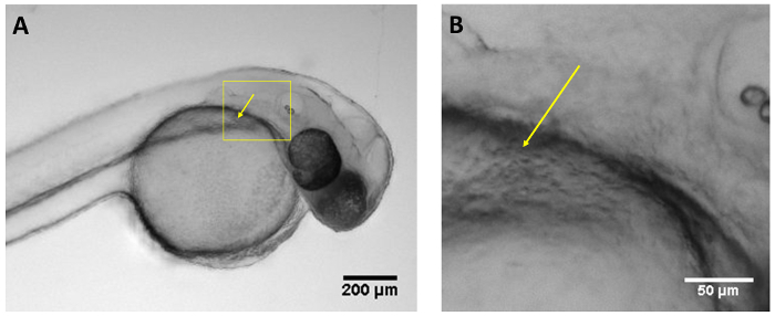
Figure 1: Optimal injection site. (A) Image of larva 48 hpf showing the optimal injection site at the duct of Cuvier (yellow arrow) using a regular stereoscope. (B) Magnified view of box in A showing the duct of Cuvier (yellow arrow). Scale bar = 200 µm (A), 50 µm (B). Please click here to view a larger version of this figure.
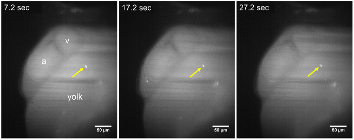
Figure 2: LSFM images of a static parasite in a 48 hpf larva. The T. cruzi parasite (yellow arrow) remains adhered to the walls of the yolk sac, throughout the time-lapse sequence (7.2 s, 17.2 s, and 27.2 s), about ~15 min after parasite injection. No change in position of the parasite is observed during an acquisition period of at least 30 s. a, Atrium; v, Ventricle. Scale bar = 50 µm. Please click here to view a larger version of this figure.
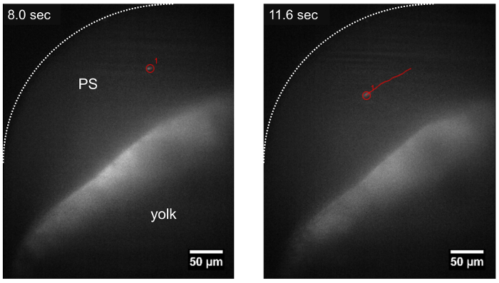
Figure 3: Trajectory of a parasite traveling in the pericardial space using LSFM. The T. cruzi parasite can be tracked while drifting in the pericardial space (PS), following the direction of blood flow (track shown in red) about ~15 min after parasite injection. Scale bar = 50 µm. Please click here to view a larger version of this figure.
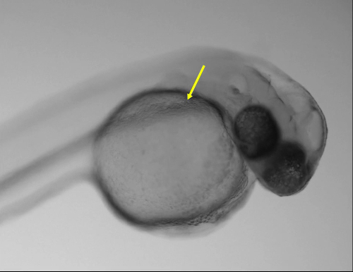
Movie 1: Blood circulation valley of the yolk in a larva 48 hpf. Movie of a larva 48 hpf showing the blood circulation valley or duct of Cuvier using a regular stereoscope. Different regions are focused during the video to show red blood cells circulating throughout the duct. Yellow arrow shows the optimal injection site. Movie was recorded about 10-15 min after parasite injection. Please click here to download the video.
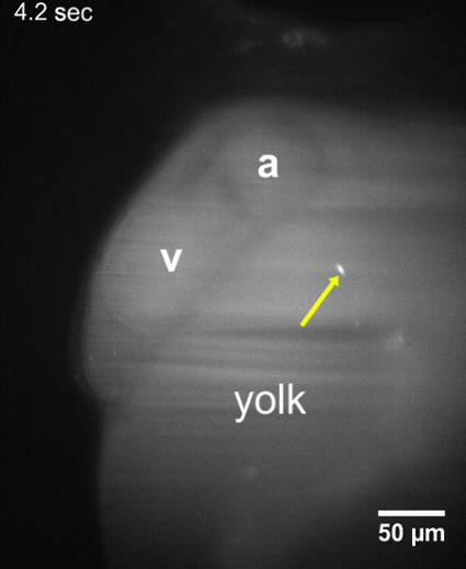
Movie 2: T. cruzi parasites attached to walls of the yolk sac. LSFM movie of a larva 48 hpf showing that T. cruzi parasites remain adhered to the yolk sac about 10-15 min after parasite injection. No change in the position of the parasite was observed during an acquisition period of at least 30 s. a, Atrium; v, Ventricle. Please click here to download the video.
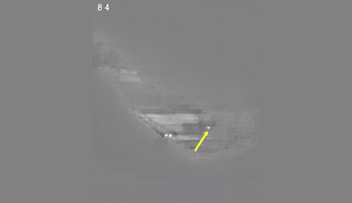
Movie 3: T. cruzi parasites attached to the walls of the heart. LSFM movie of a larva 48 hpf showing that T. cruzi parasites remain adhered to the cardiovascular wall, despite the strong heart contractions about 10-15 min after parasite injection. Erythrocytes can be observed as black rounded shadows. Please click here to download the video.
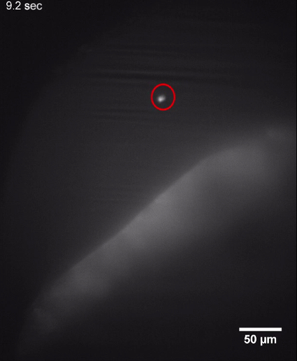
Movie 4: Parasites moving in the pericardial space. LSFM movie of a larva 48 hpf showing T. cruzi parasites drifting in the pericardial space. Two parasites can be traced at different time points (ID 1, in red circle, and ID 2 in yellow circle), following a similar trajectory. The movie was recorded about 10-15 min after parasite injection. Please click here to download the video.
Supplemental Figure 1: Accumulation of CFSE fluorescent signal in the yolk. Stereoscope images of a wildtype larva injected at 48 hpf at the duct of Cuvier. CFSE fluorescent signal progressively accumulates in the yolk, after two days post injection (48 hpi). Scale bar = 500 µm. Please click here to download the figure.
Supplemental Movie 1: Parasites attached to walls and valves of the circulatory system. Stereoscope time series images of a wild type larva injected at 48 hpf. Images are taken at 0.2 s intervals capturing T. cruzi parasites moving in synchrony with cardiac muscle contractions at the atrioventricular valve. Movie was recorded about 30 min after parasite injection. Please click here to download the video.
Supplemental Movie 2: Moving and adhered parasites in the periventricular space and yolk. LSFM movie of a larva 48 hpf showing T. cruzi parasites drifting or adhered to the pericardial space or the yolk. A transmitted light view was observed for the first 5 s. The fluorescence view was observed from 5.2-25.8 s. The movie was recorded about 10-15 min after parasite injection. Please click here to download the video.
Discussion
This study highlights the advantages of zebrafish as an animal model for studying pathogen behavior in vivo. In particular, this study proposes a method to visualize the pathogen T. cruzi in its natural environment: hematic circulation. The environment of the circulatory microenvironment in fish is comparable to that of mammals, and trypanosomatids have evolved mechanisms for traveling, evading the immune system, and attaching to cells for infection in that environment25. This protocol offers a description of an optimal procedure for culture of T. cruzi in a human cell line and subsequent isolation of flagellar forms for fluorescent labeling. It then shows the appropriate settings for successful injection of the parasites into transparent zebrafish for later mounting and visualization using LSFM. Finally, this protocol offers suggestions for efficient and effective in vivo imaging of parasite location and movement in circulation using LSFM.
The flagellum of trypanosomes emerges from its posterior region, flowing from the cell body, and hangs free at the anterior part of the organism26. T. cruzi propels itself by waving the flagellum ahead of the body, which undulates the parasite's entire body. Flagellar movement is not only indispensable for parasite motility, as in the case of T. brucei27 (the causative agent of African trypanosomiasis), but it is also used for cellular infection, as has been demonstrated in T. cruzi5,28. Although zebrafish are not the natural host for T. cruzi, the parasite's motility can be studied in a developing cardiovascular circulation system using the protocols described here. Additionally, there are trypanosomes species that infect cyprinids, the class of zebrafish, such as T. carassii and T. borreli25. These parasite species could be used to study in real time the movements and attachment mechanisms of these trypanosomatids; such studies can lend insight into the mammalian cell infection process.
In this study, injected motile T. cruzi parasites were observed traveling through the cardiovascular circulation of inoculated fish, moving along with opaque erythrocytes, and adhering to structures in the cardiovascular system walls. We used a home-built LSFM with a 10X achromatic long working distance air objective (17.6 mm) for the illumination arm with a numerical aperture of 0.25. A 40X apochromatic water immersion objective with a numerical aperture of 0.8 and a working distance of 3.5 mm was used for the detection arm. The detection objective was immersed in the sample chamber, while the illumination objective was outside the chamber. A port in the chamber sealed with a coverslip and optical glue allowed for the illumination beam to enter the chamber, as depicted in Lorenzo et al.18 For illumination, we used a 50 mW DPSS laser at 488 nm whose power was modulated by an Acousto Optical Tunable Filter. The detection path used filters compatible with Green Fluorescent Protein (GFP) or FITC. A light sheet microscope equipped with a capillary sample holder (ideally with automated rotation) and temperature control of the sample chamber should be suitable for this application. The microscope should be aligned and calibrated according to the manufacturer's instructions or user's laboratory standard protocols before acquisition, if necessary. In this protocol, we controlled the microscope using the SPIM software19.
It is important to note that in the circulation of zebrafish, larvae parasitic attachment is effective. In the cardinal vein, parasites remained attached for up to several minutes; in the heart, they held on to valves and walls, oscillating with heart contractions. Further studies remain in order to elucidate whether T. cruzi interacts with the flowing erythrocytes that drift in the direction of blood flow. Previous in vitro studies have shown that the presence of solid structures (i.e., blood cells), or increased viscosity of the liquid to mimic blood in vitro, has a significant effect on the motility and velocity of the parasite9.
There are many questions regarding the course of infection of T. cruzi in humans after amastigotes escape phagocytic cells, the initially infected cell type29. For example, how do they arrive to their target organs? What are the mechanisms for tropism to the preferred organs, such as cardiac, digestive, and central nervous systems? Interestingly, in this study the parasites were initially imaged in the heart because it was the site of highest density of parasites. However, the CFSE signal subsequently accumulated in the developing intestine by 7 days post injection. Although the anatomy of fish and mammals is different, the results of this study demonstrate a form of tropism, as it was observed that parasites exhibited tropism toward known preferred target organs despite organismal differences. One significant limitation of this study concerns the temperature used in the experiments. Zebrafish larvae should be kept around 28 °C during the entire procedure. Though this temperature might be similar to the vector host (insects of the Triatominae subfamily), it is quite different from the warm-blooded mammals that comprise the final hosts (around 37 °C). T. cruzi is known to have flagellar living forms in both hosts; however, it is important to bear in mind that this factor might have an effect in the behavior of the animal in vivo.
Although the fish´s adaptive immune system is not mature until 4 weeks post fertilization, the innate immune system is active early in development10. As early as 48 or 96 hpf, phagocytic cells were observed having engulfed labeled trypanosomes (data not shown). This limits the window of time for visualization of the parasite. However, if a study was to focus on evaluating the fish´s immune response, injection at later stages may be recommended. Also, injection of parasites into transgenic fish lines with labeled macrophages or other cells of the immune system can be useful in studying parasite attachment and possible endocytosis mechanisms. It is important to note that if the parasites are labeled with CFSE, transgenic cell labels should not be GFP, and a marker in the yellow or red end of the spectrum is required.
To assess the detailed direction of parasite movement, it may be useful to follow their trajectory in 3 dimensions (3D). For 3D visualization and reconstruction of the process, a high-speed system is necessary. With the equipment used in this protocol, it is only possible to visualize the parasites in one plane. In this case, we prioritized maintaining focal plane stability during the parasite movement, and recording its trajectory in one plane.
The methodology proposed here paves the way to further investigate parasite behavior in cardiovascular circulation. In summary, the essential steps to imaging live fluorescent parasites inside zebrafish larvae are: (i) use of early hatched embryos (24-48 hpf) or larvae, or animals between 72-96 hpf with no pigmentation so that they are transparent and easy to inject; (ii) image larvae as soon as possible after injection to avoid parasite clearance by phagocytic cells; and (iii) focus the LSFM on the site of interest (e.g., pericardial region) and maintain the focus. This novel procedure allows the visualization of trypomastigotes in an environment comparable to its natural infection niche, providing for the first time the possibility to study T. cruzi in a living organism.
Disclosures
The authors have nothing to disclose.
Acknowledgements
This work was supported by the Convocatoria Interfacultades from Vicerrectoría de Investigaciones de la Universidad de los Andes, and the USAID Research and Innovation Fellowship program. We thank Juan Rafael Buitrago and Yeferzon Ardila for fish maintenance and assistance.
Materials
| 0.5-10 μL Micropipette | Fisherbrand | 21-377-815 | |
| Agarose RA | Amresco | N605 | Regular |
| Agarose SFR | Amresco | J234 | Low Melting point |
| Aquarium salt | Instant ocean | SS15-10 | |
| Cell Count chamber | Boeco | Neubauer | |
| Cell culture flasks | Corning | 430639 | |
| Centrifuge | Sorvall | Legend RT | |
| CFSE | ThermoFisher | C34554 | |
| Detection objective | Nikon 40x 0.8NA | 40x CFI APO NIR | |
| DMEM medium | Sigma-Aldrich | D5648 | |
| Dumont #5 fine forceps | World precision Instruments | ||
| Ethyl 3-aminobenzoate methanesulfonate salt (Tricaine) | Sigma-Aldrich | A5040 | |
| Fetal calf serum (FCS) | Eurobio | CVFSVF00-01 | |
| Filter | Chroma | ET-525/50M | |
| Glass capillaries for embryo mounting | Vitrez Medical | 160215 | |
| Glass capillaries for pulling needles | World precision instruments | TW100-4 | |
| Glucose | Gibco | A2494001 | |
| HEPES | Gibco | 156300-80 | |
| Incubator | Thermo Corporation | Revco | |
| Larval microinjection mold | Adaptive Science Tools | I-34 | |
| Laser | Crystalaser | DL488-050 | |
| L-glutamine | Gibco | 250300-81 | |
| Methylene blue | Albor Químicos | 12223 | |
| Micromanipulator | Narishige | MN-153 | |
| Micromanipulator system | Sutter Instrument | MP-200 | For LSFM |
| Micropipette puller device | Narishige | PC-10 | |
| Microscope | Olympus | CX31 | |
| Microscope (inverted) | Olympus | CKX41 | |
| Multipurpose microscope | Nikon | AZ100M | |
| Neubauer counting chamber | Boeco Germany | ||
| Penicillin-streptomycin | Gibco | 15140-163 | |
| Petri dish 94×16 | Greiner bio-one | 633181 | |
| Plastic pasteur pipette | Fisherbrand | 11577722 | |
| Rotation stage | Newport | CONEX-PR50CC | |
| RPMI-1640 medium | Sigma-Aldrich | R4130 | |
| Sodium pyruvate | Gibco | 11360-070 | |
| Stereoscope | Nikon | C-LEDS | |
| Tricaine (MS-222) | Sigma-Aldrich | 886-86-2 | |
| TRIS | Amresco | M151 | |
| Trypsin-EDTA (0.25%) | Gibco | R-001-100 | |
| Tubes 15 ml | Corning | 05-527-90 |
References
- Bern, C. Chagas’s Disease. New England Journal of Medicine. 373 (5), 456-466 (2015).
- Sosa Estani, S., Segura, E. L., Ruiz, A. M., Velazquez, E., Porcel, B. M., Yampotis, C. Efficacy of chemotherapy with benznidazole in children in the indeterminate phase of Chagas’ disease. The American Journal of Tropical Medicine and Hygiene. 59 (4), 526-529 (1998).
- Rosas, F., Roa, N., Cucunuba, Z. M., Cuellar, A., Gonzalez, J. M., Puerta, J. C. Chagasic Cardiomyopathy. Cardiomyopathies – From Basic Research to Clinical Management. , (2012).
- Finkelsztein, E. J., et al. Altering the motility of Trypanosoma cruzi with rabbit polyclonal anti-peptide antibodies reduces infection to susceptible mammalian cells. Experimental Parasitology. 150, 36-43 (2015).
- Martins, R. M., Covarrubias, C., Rojas, R. G., Silber, A. M., Yoshida, N. Use of L-Proline and ATP Production by Trypanosoma cruzi Metacyclic Forms as Requirements for Host Cell Invasion. Infection and Immunity. 77 (7), 3023-3032 (2009).
- Engstler, M., et al. Hydrodynamic Flow-Mediated Protein Sorting on the Cell Surface of Trypanosomes. Cell. 131 (3), 505-515 (2007).
- Uppaluri, S., et al. Impact of Microscopic Motility on the Swimming Behavior of Parasites: Straighter Trypanosomes are More Directional. PLoS Computational Biology. 7 (6), e1002058 (2011).
- Heddergott, N., et al. Trypanosome Motion Represents an Adaptation to the Crowded Environment of the Vertebrate Bloodstream. PLoS Pathogens. 8 (11), e1003023 (2012).
- Gratacap, R. L., Wheeler, R. T. Utilization of zebrafish for intravital study of eukaryotic pathogen–host interactions. Developmental & Comparative Immunology. 46 (1), 108-115 (2014).
- Meijer, A. H., Spaink, H. P. Host-Pathogen Interactions Made Transparent with the Zebrafish Model. Current Drug Targets. 12 (7), 1000-1017 (2011).
- White, R. M., et al. Transparent Adult Zebrafish as a Tool for In Vivo Transplantation Analysis. Cell Stem Cell. 2 (2), 183-189 (2008).
- Huisken, J. Slicing embryos gently with laser light sheets. BioEssays. 34 (5), 406-411 (2012).
- Westerfield, M. . The zebrafish book: A guide for the laboratory use of zebrafish (Danio rerio). , (2000).
- Kimmel, C. B., Ballard, W. W., Kimmel, S. R., Ullmann, B., Schilling, T. F. Stages of embryonic development of the zebrafish. Developmental Dynamics. 203 (3), 253-310 (1995).
- Vargas-Zambrano, J. C., Lasso, P., Cuellar, A., Puerta, C. J., González, J. M. A human astrocytoma cell line is highly susceptible to infection with Trypanosoma cruzi. Memórias do Instituto Oswaldo Cruz. 108 (2), 212-219 (2013).
- Rosen, J. N., Sweeney, M. F., Mably, J. D. Microinjection of Zebrafish Embryos to Analyze Gene Function. JoVE (Journal of Visualized Experiments). (25), e1115-e1115 (2009).
- Lorenzo, C., et al. Live cell division dynamics monitoring in 3D large spheroid tumor models using light sheet microscopy. Cell Division. 6, 22 (2011).
- Edelstein, A. D., Tsuchida, M. A., Amodaj, N., Pinkard, H., Vale, R. D., Stuurman, N. Advanced methods of microscope control using μManager software. Journal of Biological Methods. 1 (2), e10 (2014).
- Weber, M., Mickoleit, M., Huisken, J. Multilayer Mounting for Long-term Light Sheet Microscopy of Zebrafish. Journal of Visualized Experiments. (84), (2014).
- Schindelin, J., et al. Fiji: an open-source platform for biological-image analysis. Nature Methods. 9 (7), 676-682 (2012).
- Ng, A. N. Y., et al. Formation of the digestive system in zebrafish: III. Intestinal epithelium morphogenesis. Developmental Biology. 286 (1), 114-135 (2005).
- Higuchi, M. D. L., et al. Correlation between Trypanosoma cruzi parasitism and myocardial inflammatory infiltrate in human chronic chagasic myocarditis: Light microscopy and immunohistochemical findings. Cardiovascular Pathology: The Official Journal of the Society for Cardiovascular Pathology. 2 (2), 101-106 (1993).
- Vago, A. R., Silva, D. M., Adad, S. J., Correa-Oliveira, R., d’Avila Reis, D. Chronic Chagas disease: presence of parasite DNA in the oesophagus of patients without megaoesophagus. Transactions of the Royal Society of Tropical Medicine and Hygiene. 97 (3), 308-309 (2003).
- Wiegertjes, G. F., Forlenza, M., Joerink, M., Scharsack, J. P. Parasite infections revisited. Developmental & Comparative Immunology. 29 (9), 749-758 (2005).
- De Souza, W. Basic cell biology of Trypanosoma cruzi. Current Pharmaceutical Design. 8 (4), 269-285 (2002).
- Langousis, G., Hill, K. L. Motility and more: the flagellum of Trypanosoma brucei. Nature Reviews. Microbiology. 12 (7), 505-518 (2014).
- Johnson, C. A., et al. Cellular response to Trypanosoma cruzi infection induces secretion of defensin α-1, which damages the flagellum, neutralizes trypanosome motility, and inhibits infection. Infection and Immunity. 81 (11), 4139-4148 (2013).
- Magalhães, L. M. D., Viana, A., Chiari, E., Galvão, L. M. C., Gollob, K. J., Dutra, W. O. Differential Activation of Human Monocytes and Lymphocytes by Distinct Strains of Trypanosoma cruzi. PLoS neglected tropical diseases. 9 (7), e0003816 (2015).

