Intranasal Immunization and Milk Collection in Studies of Maternal Immunization in New Zealand White Rabbits (Oryctolagus cuniculus)
Summary
This article describes and demonstrates the administration of intranasal vaccines and the collection of milk from lactating rabbits (Oryctolagus cuniculus) as a means to assess mucosal immunity in a translationally appropriate model of maternal immunization.
Abstract
Due to similarities in placentation and antibody transfer with humans, rabbits are an excellent model of maternal immunization. Additional advantages of this research model are the ease of breeding and sample collection, relatively short gestation period, and large litter sizes. Commonly assessed routes of immunization include subcutaneous, intramuscular, intranasal, and intradermal. Nonterminal sample collection for the chronological detection of the immunologic responses to these immunizations include the collection of blood, from both dams and kits, and milk from the lactating does. In this article, we will demonstrate techniques our lab has utilized in studies of maternal immunization in New Zealand White rabbits (Oryctolagus cuniculus), including intranasal immunization and milk collection.
Introduction
Studies of maternal immunization and antibody transfer are invaluable for numerous reasons, as this is the initial route of immunity transfer and subsequent protection from pathogens and diseases in newborns and infants. Maternal immunization has the potential to positively impact both maternal and infant/child health at the global level by reducing morbidity and mortality associated with certain pathogens during this vulnerable period1. The main goal of this strategy is to increase the levels of specific maternal antibodies throughout pregnancy. These antibodies can then be transferred to the newborn and infant at levels sufficient enough to protect against infections until their immune system is mature enough to adequately respond to challenges1,2,3. Previous work has demonstrated that higher antibody titers at birth are associated with either complete protection or a delayed onset and reduced severity of numerous different infectious diseases in the newborn, including tetanus, pertussis, respiratory syncytial virus (RSV), influenza, and group B streptococcal infections1,2,3.
In humans, maternal antibodies are transferred passively across the placenta and are also transferred through the breast milk via nursing. Previous work has demonstrated that HIV-specific IgA levels in human breast milk from mothers infected with the virus were associated with reduced postnatal transmission of the virus, suggesting a protective role for breast milk anti-HIV IgA4. Studies in nonhuman primates have demonstrated that immunization against HIV can induce a significant antibody response in the breast milk, and although similar serum IgG responses were induced following systemic versus mucosal immunization, mucosal immunization induced a significantly higher IgA response within the milk5,6.
Identifying a translationally appropriate animal model for these studies should take into account the placentation type and mechanisms of passive antibody transfer, as well as the transfer of antibodies through breast milk. There are three main types of placentation in mammals based on the tissue types and layers at the materno-fetal interface, including hemochorial (primates, rodents and rabbits), endotheliochorial (carnivores), and epitheliochorial (horses, pigs, and ruminants). The hemochorial placenta is the most invasive type, allowing for direct communication between the maternal blood supply and the chorion, or the outermost fetal membrane. Based on the number of trophoblast layers, there are several variations of hemochorial placentation, including the hemomonochorial placenta found in primates, the hemodichorial placenta in rabbits, and the hemotrichorial placenta observed in rats and mice7. This direct contact between maternal blood supply and chorion allows for the passive transfer of antibodies across the placenta during gestation. IgG is the only antibody class that significantly crosses the human placenta8, whereas IgA is the predominant class of Ig found in human breast milk9. Of the scientifically relevant models, only primates (including humans), rabbits, and guinea pigs transfer IgG in utero and IgA in the milk10,11. Therefore, the rabbit model incorporates factors comparable to those in humans that control transplacental transfer of IgG and lactational transfer of IgA.
In addition to serving as an exceptional model for maternal immunity and vaccine development, similarities between the rabbit and human nasal cavities make them an appropriate model for intranasal immunization. The volume of the rabbit nasal cavity is more similar to humans than rodent models based on relative body mass12. Additionally, Casteleyn et al. 12 demonstrated that the nasal associated lymphoid tissue (NALT) is more voluminous in the rabbit compared to rodents. The NALT is located primarily at the ventral and ventromedial aspect of the ventral nasal meatus and at the lateral and dorsolateral aspect of the nasopharyngeal meatus in rabbits, whereas in rodents, the lymphoid tissue is only present along the ventral aspect of the nasopharyngeal meatus12. In rabbits, the structure and location of the intraepithelial and lamina propria lymphocytes, as well as the isolated lymphoid follicles, are similar to humans12.
Additional advantages of using the rabbit as a model for maternal and mucosal immunity include their high fecundity and relatively short gestation period. Large auricular blood vessels allow for relatively easy access to large volumes of blood for serial collections. A variety of mucosal samples can be collected for antigen-specific antibody response assays, including breast milk13 (when lactating), mucosal secretions or washes (e.g., oral14,15,16, bronchoalveolar lavage13,17,18,19, vaginal20,21,22), and feces20,23,24,25. Milk samples can be easily collected during lactation to assess the presence of antigen-specific antibody responses. Though not as abundant as for humans and mice, a wide variety of experimental reagents are available for rabbit-specific studies and assays. In this article, we will describe and demonstrate intranasal immunization and milk collection in New Zealand White rabbits (Oryctolagus cuniculus).
Protocol
All procedures were approved and performed in accordance with the Duke University IACUC policies.
NOTE: Materials needed are provided in the Table of Materials.
1. Rabbit Sedation and Anesthesia
- Sedate the female rabbit (sexually mature; approximately 5-30 months old) by administering acepromazine intramuscularly (IM) at a dose of 1 mg/kg. Depending on the size of the animal, use a 1 or 3 mL syringe with a 25 G needle. Epaxial muscles are the preferred site of the intramuscular injection.
NOTE: Acepromazine can also be administered subcutaneously, but IM is preferred by the lab, as it acts more rapidly and reduces the incidence of skin lesions. - Wait 10-15 minutes to allow the acepromazine to take effect.
- Anesthetize the rabbit with isoflurane by placing the connected nose cone over the animal's nose. Adjust the vaporizer to up to 4% isoflurane combined with up to 4 liters/minute oxygen. Rabbits have a high aversion to isoflurane, so adequate restraint is necessary when masking the animal.
- Once fully anesthetized, as assessed by the pinna, pedal, and/or palpebral reflex, apply ophthalmic lubricant to each eye to prevent drying of the eyes and subsequent corneal ulceration.
- Continually monitor reflexes and breathing during anesthesia, and reduce the isoflurane rate to 1-2% once an adequate plane of anesthesia has been reached.
2. Intranasal immunization
- Prepare immunization solution prior to animal handling.
- Sedate the rabbit as described above.
- Once the lab member is ready to administer the vaccine and the rabbit is in an adequate plane of anesthesia, turn off the isoflurane and oxygen and remove the nose cone.
- Place the rabbit in dorsal recumbency, and prop the neck and head at an approximate 45° angle that allows easy access and visualization of both nares by the lab member administering the vaccine.
- Load the pipette with no more than 100 µL of the vaccine solution, and quickly administer the solution in each nostril. The pipette should be held at an approximate 45° angle, angled towards the medial aspect of the nasal passage.
NOTE: The goal of immunization is for the solution to contact the mucosal membrane of the nares, so the tip should not be placed within the nares, as this may result in abrasion or irritation of the mucosal tissues and potentially influence the immunogenicity of the nasally-administered vaccine. The vaccine should be administered quickly and done in the same manner in the other nare. - Following administration in both nasal passages, maintain the rabbit in dorsal recumbency for 30 seconds to minimize leakage of the vaccine solution.
NOTE: The lab will typically administer no more than 100 µL per nostril at a time. If a larger volume is to be administered, with a maximum total of 500 µL, the vaccine can be given in 100 µL aliquots with a 30 second rest period between immunizations, and additional administrations of vaccine repeated, with 30 seconds of rest between each administration, until the total vaccine volume is delivered. - Following immunization, place the rabbit on the ventrum for recovery and closely monitor the animal until it can maintain sternal recumbency.
3. Milk collection
- Sedate the lactating rabbit as described above.
- Clean the skin over the marginal ear vein with the alcohol swab/wipe.
- Using a 1 mL syringe and 25 g needle, administer approximately 1-2 IU of oxytocin intravenously via the marginal ear vein to induce milk letdown.
NOTE: Due to the smooth muscle relaxation, it is common for the rabbit to urinate or defecate following administration of oxytocin. - Following oxytocin administration, apply pressure to the injection site with the piece of gauze.
- While maintaining the anesthesia mask over the rabbit's nose, prop the rabbit on its hindquarters.
NOTE: Milk collection can also be performed with the animal in lateral recumbency, but the lab finds that collection is easier when the rabbit is propped up on its rump with an assistant holding the rabbit upright with the anesthesia mask. - Open the sterile tube to prepare for milk collection and locate the mammary tissue and associated teats. The teats are typically surrounded by wet fur from recent nursing, and the mammary tissue is easily palpable when full of milk.
- Grasp the mammary tissue associated with a teat between the thumb and forefinger and apply a gentle, massaging pressure to the glandular tissue in the direction of the teat. Place the collection tube over the teat to collect the expressed milk.
NOTE: It can sometimes take several minutes for the oxytocin to be effective, and milk production appears to vary among mammary glands. If milk expression is not successful, wait several minutes or rotate around to the additional mammary glands. Milk from all teats can be collected in the same vial. Typically, several milliliters of milk can be easily collected from a lactating doe. - Following milk collection, turn the isoflurane and oxygen off, and allow the rabbit to recover while being closely monitored until the animal is able to maintain sternal recumbency.
Representative Results
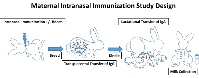
An overview of a typical maternal intranasal immunization study design is depicted in Figure 1, incorporating the immunizations, breeding, kindling, lactation, and antibody transfer. Though not illustrated, blood should be collected prior to the initial immunization for baseline measurements and throughout the remainder of the study at regular intervals. Blood is easily obtained via the central ear artery with mild sedation and a topical analgesic agent (e.g., lidocaine 2.5% and prilocaine 2.5% cream). The presence of antigen-specific IgG levels can be measured in these samples. Female rabbits are immunized via the intranasal route, as described in the protocol and demonstrated in the video. Depending on the study, the vaccine may require a boost or may need to be given through an additional route (e.g., intramuscular or subcutaneous). Following study initiation, rabbits are bred; we prefer to purchase proven breeders from vendors to use to ensure a higher pregnancy rate for these studies. Depending on the immunization timeline, rabbits may receive additional immunizations throughout pregnancy. Antigen-specific IgG is transferred transplacentally to the kits, and at approximately 30-32 days post-breeding, pregnant does will kindle. We recommend limiting handling of kits for the first several days to minimize rejection from the does. Blood samples can be collected from the kits to assess antigen-specific IgG levels that were transferred transplacentally (Figure 3). In addition to a wide variety of nutrients, kits receive IgA from the lactating doe while nursing. Kits are typically weaned at 4-8 weeks, but prior to weaning, milk can easily be collected from lactating does, as demonstrated in the video. The collected milk samples can then be processed for the detection of total and antigen-specific IgA levels (Figure 4). Depending on the study, vaccines (+/- boosts) can be administered to the kits, and serial blood samples can be collected from the kits at a very early age using the lateral saphenous vein.
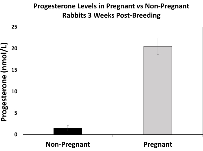
For maternal studies, determining pregnancy as early as possible is helpful for the study design and for ensuring the doe does not need to be rebred. Progesterone measurements can be used as a means to detect pregnancy. As shown in Figure 2, elevated progesterone levels can be detected in pregnant rabbits compared to non-pregnant rabbits even after matings by a buck were confirmed for all does. There are additional methods for pregnancy detection, including manual palpation, ultrasound, and radiographs; however, these require well-trained personal and proper equipment.
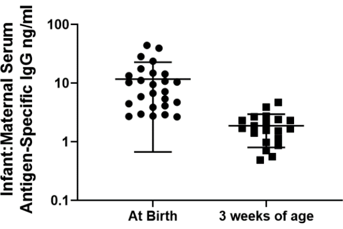
Antigen-specific IgG that was transferred transplacentally while in utero can be measured in the serum of kits. Blood can be collected from a small number of kits at or near the time of birth to assess early antigen-specific antibody levels, but serial blood collection is technically much easier as the kits age and increase is size. As depicted in Figure 3, serum levels of antigen-specific IgG in the kits can be measured by ELISA and compared to the maternal levels. Maternally transferred IgG levels tend to be higher at birth and decrease over time.
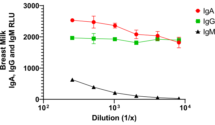
As a type of mucosal sample, milk can be collected and processed to measure total or antigen-specific antibody levels. As shown in Figure 4, IgA makes up a significant portion of the total antibody levels within the breast milk that is being transferred to the kits via lactation. Our results demonstrate that breast milk IgA produces a slightly higher ELISA signal (relative light units, RLU) when compared to IgG, and both IgA and IgG produce a signal that is much higher than the signal for IgM. These results are in agreement with results from others that suggest that rabbit milk contains around 4.5 mg/mL IgA, 2.4 mg/mL IgG, and 0.1 mg/mL IgM26,27.

Figure 1. Sample timeline for a maternal intranasal immunization study design in a rabbit (Oryctolagus cuniculus) model. Female rabbits are immunized via the intranasal route, as described in the protocol and demonstrated in the video. Depending on the study, the vaccine may require a boost or may need to be given through an additional route (e.g. intramuscular or subcutaneous). Rabbits are then bred. Antigen-specific IgG is transferred transplacentally to the kits, and at approximately 30-32 days post-breeding, pregnant does will kindle. IgA is passed to the kits from the lactating doe while nursing. Prior to weaning, milk can easily be collected from the lactating does to assess total and antigen-specific IgA levels. Please click here to view a larger version of this figure.

Figure 2. Progesterone levels in pregnant and non-pregnant rabbits at 3 weeks post-breeding. Blood was collected from rabbits at 3 weeks post-breeding. Rabbits were confirmed either pregnant or non-pregnant based on ability to kindle a litter at 30-32 days post-breeding. Serum progesterone levels were measured through the Michigan State University Veterinary Diagnostic Laboratory using a chemiluminescent immunoassay (CLIA) with an immunoassay system (e.g., Siemens Healthineers IMMULITE 2000). Error bars represent standard error of the mean, and the sample size consisted of 4-6 rabbits per group. Please click here to view a larger version of this figure.

Figure 3. Antigen-specific IgG levels in kits (relative to maternal levels) at birth and at 3 weeks of age following a series of maternal immunizations. Blood was collected from the does and kits soon after kindling and at 3 weeks of age. Antigen-specific IgG levels within the serum were detected using a fluorescent ELISA as previously described28. Antigen-specific IgG is plotted as a ratio of levels detected in the kit serum and maternal serum. Please click here to view a larger version of this figure.

Figure 4. A comparison of IgA, IgM, and IgG levels in rabbit milk. Rabbit milk was collected as described and demonstrated in the video. Milk was processed by a long centrifugation (13,000 x g for 4.5 hours at 4 °C), and the clear middle layer was isolated following processing. Total IgA, IgG, and IgM levels were measured in this clear layer by fluorescent ELISA as previously described28, except that plates were coated with polyclonal anti-IgA, anti-IgG, or anti-IgM to detect total rabbit IgA, IgG, or IgM, respectively. Please click here to view a larger version of this figure.
Discussion
Although not described in the above protocol, successful breeding of the rabbits is necessary for this maternal model and to allow for milk collection. Rabbits are easily bred by live cover in a research setting. It is recommended that does be transferred to the buck's cage for breeding, as does can be territorial and aggressive if kept in their own cage with the buck. If females are non-receptive after 15 minutes (as indicated by running away biting, or vocalizing), the doe should be placed back into her own cage. There are several informative videos and tutorials on rabbit breeding that can be observed online29, but after breeding, the male will typically fall over and may vocalize. Once this is observed, the doe can be returned to her cage. A buck may breed 2-3 times a day with no decrease in sperm count30. In our IACUC-approved protocol, bucks are limited to breeding 10-12 does per week and are provided at least two rest days per week, as sources state that a single buck is usually sufficient to service 10-15 does31. Our group suggests purchasing proven breeders from the vendor to improve the breeding success rate. As rabbits are induced ovulators and ovulation typically occurs 10-13 hours post-copulation31, we have experienced a higher rate of pregnancy in does that are bred in the morning and then again in the afternoon, or twice within that 10-13 hour window (observed increase from 75% to 95% success rate, unpublished). Based on the literature, typical breeding success rates vary from 57-100%32,33,34,35,36,37,38,39,40, and litter sizes average approximately 7-9 kits31.
Determining pregnancy as early as possible is helpful in maternal studies to confirm that the doe does not need to be rebred or removed from the study. Options for pregnancy detection include palpation (as early as 14 days)31, ultrasound (as early as 5-9 days)40, radiographs (as early as day 11)31, weight gain, and molecular techniques, such as measurements of insulin-growth factors41 (IGF) and progesterone34,37,38,42,43. Previous work has indicated significant elevations of IGF-II levels in pregnant rabbits compared to levels in non-pregnant rabbits41. However, in our hands, we were unable to detect a difference in IGF-II levels between pregnant and non-pregnant rabbits (unpublished). As adequate progesterone levels are necessary for the maintenance of pregnancy in rabbits37,44, several studies have assessed progesterone levels in pregnant rabbits and demonstrated elevated levels relative to non-pregnant rabbits, particularly during organogenesis around mid-gestation34,43,44. Our group was unable to detect differences in progesterone levels using an ELISA between pregnant and non-pregnant rabbits, but preliminary results using an automated chemiluminescent assay at the Michigan State University Veterinary Diagnostic Laboratory indicate elevated progesterone levels in pregnant rabbits compared to non-pregnant rabbits even after matings by a buck were confirmed for all does assessed (Figure 2).
Lactating does can produce approximately 250 mL, or 60 mL/kg, of milk daily45,46, allowing for large volumes for experimental assays to assess total and antigen-specific antibody responses/concentrations. Rabbit milk contains high levels of fat and protein, containing 2 and 3 times more concentrated levels of fat and protein, as compared to cow and sow milk, respectively45,47. Due to the high fat content in the milk, the samples require significant processing, depending on the assays to be conducted. Following centrifugation of the milk sample, three distinct layers are separated out, including the cells within the bottom layer, clear intermediate layer containing the immunoglobulins, and fat within the top layer48. Immunoglobulins, within the clear intermediate layer, are present at high concentrations within the colostrum and milk and are primarily made up of IgA, IgG, and IgM (Figure 4). Though milk samples are easily collected by hand, and this is our preferred technique within the lab, vacuum systems have also been reported in the literature13,46,48. Yoshiyama et al.48 collected milk samples using negative pressure and described a long centrifugation (15,000 x g for 4 hours) for separation of the milk layers prior to passing the clear intermediate layer through a Sepharose 4B column for immunoglobulin removal. Using this method, authors were able to detect cholera toxin-specific antibodies within the rabbit milk of orally immunized rabbits at sufficient levels for protection against Vibrio cholerae-induced secretion in the intestine48. Peri et al.13 processed milk samples collected by aspiration with a water vacuum system by centrifugation at 4 °C for 2 hours at 24,000 x g. In this study, authors were able to detect anti-RSV IgA in the colostrum, milk, and bronchial and intestinal secretions following mucosal immunization (oral or intratracheal), but not intravenous immunization, whereas anti-RSV IgG was detected in colostrum, milk and serum regardless of immunization route13.
Care should be taken during the intranasal immunization procedure to confirm the rabbit is in an adequate plane of anesthesia and to avoid administering large volumes of vaccine at once. Previous work from our group has shown that the efficacy of the intranasal immunization was influenced by the use of anesthesia49. Specifically, deep anesthesia induced by the combination of ketamine and xylazine correlated with increased immunogenicity of the nasally administered vaccine and improved nasal retention of the solution. These levels were significantly higher than retention and immunogenicity in rabbits intranasally immunized following sedation with acepromazine and butorphanol49. Similar findings have also been demonstrated following intranasal immunization in mice50,51. Additionally, our work demonstrated increased immunogenicity following intranasal immunization under the deep anesthesia over animals that were fully awake and over animals undergoing shorter duration anesthesia with a combination of acepromazine and isoflurane. This difference in IgG immunogenicity between deep versus shorter duration anesthesia was significant at the 42 day time point but not at the 56 day time point. Although increased immunogenicity and retention may result from the deeper anesthesia, rabbits required at least 30 minutes to recover; whereas, rabbits were upright and mobile within 5 minutes of receiving the nasal vaccine following the shorter duration isoflurane-induced anesthesia. Although anesthesia may not be ideal for mimicking the clinical setting for intranasal immunization in humans, a shorter duration anesthesia (e.g., isoflurane) may be preferred to a longer, deep anesthesia with injectable agents (e.g., ketamine + xylazine). The use of an animal model, as demonstrated in Gwinn et al.49, will often require the use of sedation or anesthesia to allow for safe animal handling and effective and consistent vaccine delivery. A potential limitation for anesthesia use with vaccine administration in animal models is its effect on the ability of the animal to induce an appropriate immune response to the vaccine. Interestingly, reports in the literature suggest that isoflurane can attenuate systemic inflammatory responses, providing protection against challenges52 and reducing oxidative stress and inflammation53.
With respect to minimization of vaccine loss and adequate intranasal delivery, our group recommends a rest period of 30 seconds after administering each aliquot of the intranasal vaccine to allow for retention of the solution within the nasal cavity and to prevent the solution from seeping out from the nostrils if the rabbit is being immediately placed in sternal recumbency and returned to the cage. Additionally, it is important to limit the volume of the aliquot administered to ensure intranasal immunization, as mucosal absorption is limited by the surface area of the nasal passage. If a large volume is administered, the solution may bypass the nasal mucosa and result in inhalational or gastric immunization. Our group recommends aliquots of 100 µL per nostril with a maximal intranasal volume of 500 µL.
This article focuses on mucosal vaccine delivery, specifically via the intranasal route, but there are multiple methods of immunization, both mucosal and parenteral that have been demonstrated in rabbits. These additional methods include, but are not limited to, oral, subcutaneous, intramuscular, and intradermal. As such, samples to be collected and assessed vary based on the experimental goals and route of immunization.
Declarações
The authors have nothing to disclose.
Acknowledgements
The authors would like to acknowledge the Division of Laboratory Animal Resources at Duke University and their husbandry team for their assistance and great care provided to the animals. Additionally, the authors would like to recognize the PhotoPath team within the Department of Pathology for their assistance with the audio and video portions of the manuscript. This work was supported by discretionary research funds from the Staats laboratory.
Materials
| Intranasal Immunization | |||
| Anesthesia Machine/Vaporizer | Vet Equip | 901807 | |
| Hypodermic Needle (25 g) | Terumo | 07-806-7584 | |
| Isoflurane (250 mL Bottle) | Patterson Veterinary | 07-893-1389 | 2-4% |
| Luer Lock Syringe (1 mL) | Air-Tite | 07-892-7410 | |
| Mucosal Vaccine | N/A | N/A | Experimental Vaccine |
| Nose Cone | McCulloch Medical | 07891-1097 | |
| Pipette Tips | VWR | 53503-290 | |
| Pipettor | VWR | 89079-962 | |
| PromAce (Acepromazine maleate) | Boehringer Ingelheim | 07-893-5734 | 1mg/kg IM |
| Puralube Sterile Ophthalmic Ointment | Dechra | 07-888-2572 | |
| Milk Collection | |||
| Alcohol Prep 2-ply | Covidien | 07-839-8871 | |
| Anesthesia Machine/Vaporizer | Vet Equip | 901807 | |
| Hypodermic Needles (25 g) | Terumo | 07-806-7584 | |
| Isoflurane (250 mL Bottle) | Patterson Veterinary | 07-893-1389 | 2-4% |
| Luer Lock Syringe (1 mL) | Air-Tite | 07-892-7410 | |
| Non-Woven Sponge (4×4) | Covidien | 07-891-5815 | |
| Nose Cone | McCulloch Medical | 07891-1097 | |
| PromAce (Acepromazine Maleate) | Boehringer Ingelheim | 07-893-5734 | 1mg/kg IM |
| Puralube Sterile Ophthalmic Ointment | Dechra | 07-888-2572 | |
| Sterile Conical Vial (15 mL) | Falcon | 14-959-49B |
Referências
- Munoz, F. M. Current Challenges and Achievements in Maternal Immunization Research. Frontiers in Immunology. 9, 436 (2018).
- Kachikis, A., Englund, J. A. Maternal immunization: Optimizing protection for the mother and infant. The Journal of Infection. 72, 83-90 (2016).
- Omer, S. B. Maternal Immunization. The New England Journal of Medicine. 376 (13), 1256-1267 (2017).
- Pollara, J., et al. Association of HIV-1 Envelope-Specific Breast Milk IgA Responses with Reduced Risk of Postnatal Mother-to-Child Transmission of HIV-1. Journal of Virology. , 01560 (2015).
- Fouda, G. G. A., et al. Mucosal Immunization of Lactating Female Rhesus Monkeys with a Transmitted/Founder HIV-1 Envelope Induces Strong Env-Specific IgA Antibody Responses in Breast Milk. Journal of Virology. 87 (12), 6986-6999 (2013).
- Nelson, C. S., et al. Combined HIV-1 Envelope Systemic and Mucosal Immunization of Lactating Rhesus Monkeys Induces Robust IgA-Isotype B Cell Response in Breast Milk. Journal of Virology. 90 (10), 4951-4965 (2016).
- Furukawa, S., Kuroda, Y., Sugiyama, A. A comparison of the histological structure of the placenta in experimental animals. Journal of Toxicologic Pathology. 27 (1), 11-18 (2014).
- Palmeira, P., Quinello, C., Silveira-Lessa, A. L., Zago, C. A., Carneiro-Sampaio, M. IgG placental transfer in healthy and pathological pregnancies. Clinical & Developmental Immunology. 2012, 985646 (2012).
- Macchiaverni, P., et al. Mother to child transfer of IgG and IgA antibodies against Dermatophagoides pteronyssinus. Scandinavian Journal of Immunology. 74 (6), 619-627 (2011).
- Pentsuk, N., vander Laan, J. W. An interspecies comparison of placental antibody transfer: new insights into developmental toxicity testing of monoclonal antibodies. Birth Defects Research Part B Developmental and Reproductive Toxicology. 86 (4), 328-344 (2009).
- Butler, J. E., Rainard, P., Lippolis, J., Salmon, H., Kacskovics, I., Mestecky, J. . Mucosal Immunology. , 2269-2306 (2015).
- Casteleyn, C., Broos, A. M., Simoens, P., Vanden Broeck, W. NALT (nasal cavity-associated lymphoid tissue) in the rabbit. Veterinary Immunology Immunopathology. 133 (2-4), 212-218 (2010).
- Peri, B. A., et al. Antibody content of rabbit milk and serum following inhalation or ingestion of respiratory syncytial virus and bovine serum albumin. Clinical and Experimental Immunology. 48 (1), 91-101 (1982).
- Fukuizumi, T., Inoue, H., Tsujisawa, T., Uchiyama, C. Tonsillar application of killed Streptococcus mutans induces specific antibodies in rabbit saliva and blood plasma without inducing a cross-reacting antibody to human cardiac muscle. Infection and Immunity. 65 (11), 4558-4563 (1997).
- Saeed, M. I., Omar, A. R., Hussein, M. Z., Elkhidir, I. M., Sekawi, Z. Development of enhanced antibody response toward dual delivery of nano-adjuvant adsorbed human Enterovirus-71 vaccine encapsulated carrier. Human Vaccines & Immunotherapeutics. 11 (10), 2414-2424 (2015).
- Ma, Y., et al. Vaccine delivery to the oral cavity using coated microneedles induces systemic and mucosal immunity. Pharmaceutical Research. 31 (9), 2393-2403 (2014).
- Jarvinen, L. Z., Hogenesch, H., Suckow, M. A., Bowersock, T. L. Induction of protective immunity in rabbits by coadministration of inactivated Pasteurella multocida toxin and potassium thiocyanate extract. Infection and Immunity. 66 (8), 3788-3795 (1998).
- Suckow, M. A., Bowersock, T. L., Nielsen, K., Grigdesby, C. F. Enhancement of respiratory immunity to Pasteurella multocida by cholera toxin in rabbits. Laboratory Animals. 30 (2), 120-126 (1996).
- Jarvinen, L. Z., HogenEsch, H., Suckow, M. A., Bowersock, T. L. Intranasal vaccination of New Zealand white rabbits against pasteurellosis, using alginate-encapsulated Pasteurella multocida toxin and potassium thiocyanate extract. Comparative Medicine. 50 (3), 263-269 (2000).
- Winchell, J. M., Routray, S., Betts, P. W., Van Kruiningen, H. J., Silbart, L. K. Mucosal and systemic antibody responses to a C4/V3 construct following DNA vaccination of rabbits via the Peyer’s patch. The Journal of Infectious Diseases. 178 (3), 850-853 (1998).
- Wegmann, F., et al. A novel strategy for inducing enhanced mucosal HIV-1 antibody responses in an anti-inflammatory environment. PLoS One. 6 (1), 15861 (2011).
- Winchell, J. M., Van Kruiningen, H. J., Silbart, L. K. Mucosal immune response to an HIV C4/V3 peptide following nasal or intestinal immunization of rabbits. AIDS Research and Human Retroviruses. 13 (10), 881-889 (1997).
- Knudsen, C., et al. Quantitative feed restriction rather than caloric restriction modulates the immune response of growing rabbits. The Journal of Nutrition. 145 (3), 483-489 (2015).
- Ciarlet, M., et al. Subunit rotavirus vaccine administered parenterally to rabbits induces active protective immunity. Journal of Virology. 72 (11), 9233-9246 (1998).
- Denchev, V., Mitov, I., Marinova, S., Linde, K. Local and systemic immune response in rabbits after intraintestinal immunization with a double-marker attenuated strain of Salmonella typhimurium. Journal of Hygiene, Epidemiology, Microbiology, and Immunology. 32 (4), 457-465 (1988).
- Watson, D. L. Immunological functions of the mammary gland and its secretion–comparative review. Australian Journal of Biological Sciences. 33 (4), 403-422 (1980).
- Lascelles, A. K., McDowell, G. H. Localized humoral immunity with particular reference to ruminants. Transplantation Reviews. 19, 170-208 (1974).
- Jones, D. I., et al. Optimized Mucosal Modified Vaccinia Virus Ankara Prime/Soluble gp120 Boost HIV Vaccination Regimen Induces Antibody Responses Similar to Those of an Intramuscular Regimen. Journal of Virology. 93 (14), (2019).
- Rabbit Tracks: Breeding Techniques and Management. Michigan State University Available from: https://www.canr.msu.edu/resources/rabit (2020)
- Patton, N. M., Manning, P. J., Ringler, D. H., Newcomer, C. E. . The Biology of the Laboratory Rabbit. , 27-45 (1994).
- Nowland, M. H., Brammer, D. W., Garcia, A., Rush, H. G., Fox, J. G., et al. . Laboratory Animal Medicine. , 411-461 (2015).
- Attia, Y. A., Al-Hanoun, A., El-Din, A. E., Bovera, F., Shewika, Y. E. Effect of bee pollen levels on productive, reproductive and blood traits of NZW rabbits. Journal of Animal Physiology and Animal Nutrition. 95 (3), 294-303 (2011).
- Feussner, E. L., Lightkep, G. E., Hennesy, R. A., Hoberman, A. M., Christian, M. S. A decade of rabbit fertility data: study of historical control animals. Teratology. 46 (4), 349-365 (1992).
- Manal, A. F., Tony, M. A., Ezzo, O. H. Feed restriction of pregnant nulliparous rabbit does: consequences on reproductive performance and maternal behaviour. Animal Reproduction Science. 120 (1-4), 179-186 (2010).
- Marai, I. F., Ayyat, M. S., Abd el-Monem, U. M. Growth performance and reproductive traits at first parity of New Zealand white female rabbits as affected by heat stress and its alleviation under Egyptian conditions. Tropical Animal Health and Production. 33 (6), 451-462 (2001).
- Rodríguez, M., et al. A diet supplemented with n-3 polyunsaturated fatty acids influences the metabomscic and endocrine response of rabbit does and their offspring. Journal of Animal Science. 95 (6), 2690-2700 (2017).
- Salem, A. A., El-Shahawy, N. A., Shabaan, H. M., Kobeisy, M. Effect of punicalagin and human chorionic gonadotropin on body weight and reproductive traits in maiden rabbit does. Veterinary and Animal Science. 10, 100140 (2020).
- Sirotkin, A. V., Parkanyi, V., Pivko, J. High temperature impairs rabbit viability, feed consumption, growth and fecundity: examination of endocrine mechanisms. Domestic Animal Endocrinology. 74, 106478 (2020).
- Sun, L., et al. Effect of light intensity on ovarian gene expression, reproductive performance and body weight of rabbit does. Animal Reproduction Science. 183, 118-125 (2017).
- El-Gayar, M., et al. Pregnancy detectuib ub rabbits by ultrasonography as compared to manual palpation. Egyptian Journal of Animal Production. 51 (3), 196-199 (2014).
- Nason, K. S., Binder, N. D., Labarta, J. I., Rosenfeld, R. G., Gargosky, S. E. IGF-II and IGF-binding proteins increase dramatically during rabbit pregnancy. The Journal of Endocrinology. 148 (1), 121-130 (1996).
- Haneda, R., et al. Changes in blood parameters in pregnant Japanese White rabbits. The Journal Toxicological Sciences. 35 (5), 773-778 (2010).
- Mizoguchi, Y., et al. Changes in blood parameters in New Zealand White rabbits during pregnancy. Laboratory Animals. 44 (1), 33-39 (2010).
- Salem, A. A., Gomaa, Y. A. Effect of combination vitamin E and single long-acting progesterone dose on enhancing pregnancy outcomes in the first two parities of young rabbit does. Animal Reproduction Science. 150 (1-2), 35-43 (2014).
- Maertens, L., Lebas, F., Szendro, Z. Rabbit milk: A review of quantity, quality and non-dietary affecting factors. World Rabbit Science. 14 (4), 205-230 (2006).
- Pekow, C. A., Suckow, M. A., Stevens, K. A., Wilson, R. P. . The Laboratory Rabbit, Guinea Pig, Hamster, and Other Rodents. , 243-258 (2012).
- Jenness, R. Lactational Performance of Various Mammalian-Species. Journal of Dairy Science. 69 (3), 869-885 (1986).
- Yoshiyama, Y., Brown, W. R. Specific antibodies to cholera toxin in rabbit milk are protective against Vibrio cholerae-induced intestinal secretion. Immunology. 61 (4), 543-547 (1987).
- Gwinn, W. M., et al. Effective induction of protective systemic immunity with nasally administered vaccines adjuvanted with IL-1. Vaccine. 28 (42), 6901-6914 (2010).
- Sloat, B. R., Cui, Z. Evaluation of the immune response induced by a nasal anthrax vaccine based on the protective antigen protein in anaesthetized and non-anaesthetized mice. Journal of Pharmcy and Pharmacology. 58 (4), 439-447 (2006).
- Janakova, L., et al. Influence of intravenous anesthesia on mucosal and systemic antibody responses to nasal vaccines. Infection and Immunity. 70 (10), 5479-5484 (2002).
- Fuentes, J. M., et al. General anesthesia delays the inflammatory response and increases survival for mice with endotoxic shock. Clinical and Vaccine Immunology. 13 (2), 281-288 (2006).
- Lee, Y. -. M., Song, B. C., Yeum, K. -. J. Impact of Volatile Anesthetics on Oxidative Stress and Inflammation. BioMed Research International. 2015, 242709 (2015).

