誘導加熱の小さな磁気圏を取り巻く水の近赤外温度計測
Summary
誘導加熱の小さな磁気圏を取り巻く水の温度を測定する 1150 と 1412 nm の波長を利用した手法である. します。
Abstract
水と周囲の電磁誘導加熱の小さな磁性球非濁った水溶液の温度を測定する手法である. します。この手法は、1150 と 1412 nm、水の吸収係数は温度に依存する波長を利用しています。水または 2.0 mm または 0.5 mm 径の磁気球を含む非混濁水系ゲル 1150 nm または選択した狭いバンドパス フィルターを使用した場合と、1412 の nm の入射光を照射します。さらに、吸収係数の横突起である吸光度の二次元画像は、近赤外カメラを介して取得されます。三次元温度分布は、球対称であると仮定することができるときに、吸光度プロファイルに逆アベルに変換を適用することによって推定します。温度は時間と誘導加熱電源によると一貫して変更する観察されました。
Introduction
媒体内の小さな熱源付近の温度を計測する技術は、多くの科学的な研究分野とアプリケーションに必要です。たとえば、磁気ハイパーサーミアの研究でがん治療法である磁性粒子の磁気小さな電磁誘導を使用して、それは磁気によって生成される温度分布を正確に予測する重要な粒子1,2。ただし、ただし、マイクロ波磁気共鳴11 、ラマン10、光音響9超音波5,6,7,83,4、12-ベース温度測定技術を研究、開発している、現時点では、このような内部の温度分布を正確に測定できません。これまで単一位置温度またはいくつかのポジションでの温度は誘導加熱の場合、非磁性光ファイバー温度センサー13,14である温度センサーで測定しました。また、メディアの表面温度は内部温度14を推定する赤外放射温度計によるリモートで観測されています。ただし、小さな熱源を含む培地が水層または非散乱体の水溶液中、私たちを示している温度15,16を測定する近赤外線 (NIR) 吸収法が役 17,18,19。本稿ではこの手法および代表的な結果の詳しいプロトコル。
近赤外吸収法は、近赤外領域での水の吸収帯の温度依存性の原理に基づいています。水は温度として 1250 nm 波長 (λ) 範囲に 1100 nm の短波長にシフト観測図 1 a、 ν1 + ν2 + のν3吸収帯にあるとおり19をが増加します。ここで、 ν1 + ν2 + このバンドは 3 つの基本的な O H 振動モードの組み合わせに対応するν3手段: 対称ストレッチ (ν1) 曲げ (ν 2)、および逆対称伸縮 (ν3)20,21。このスペクトルの変化は、バンドで最も温度敏感な波長がλ ≈ 1150 nm であることを示します。水の他の吸収バンドは、温度15,16,17,18,20,21に関して同様の動作も展示します。Ν1 + ν3バンド水の観察範囲λ以内 = 1350−1500 nm とその温度依存性を図 1 bに示します。Ν1 + 水のν3バンドで、1412 nm が最も温度に敏感な波長です。したがって、それはλで吸光度の 2 D 画像をキャプチャする近赤外カメラを用いた二次元 (2 D) 温度画像を取得する = 1150 または 1412 nm。水の吸収係数としてλ = 1150 nm は、 λ = 1412 nm よりも小さく、元の波長は約 10 mm 厚の水性メディアに適した、後者は約 1 mm 厚のものに適しています。最近では、 λを使用して = 1150 nm、直径 1 mm の鋼球を誘導加熱19を含む 10 mm 厚水層内の温度分布を得た。また、 λを使用して 0.5 mm 厚い水の層内の温度分布を測定した 1412 nm15,17を =。
イメージング法 NIR ベース温度の利点は簡単にセットアップおよび透過吸収測定法で、ない蛍光、蛍光体、または他のサーマル プローブを必要があるために、実装します。さらに、その温度分解能は 0.2 K15,17,19未満です。このような良い温度分解能は、干渉法による、熱と物質移動研究22,23,24でよく使用されている伝送方式は他では達成できません。我々 は、しかし、イメージング法 NIR ベースの温度は適していませんかなりローカル温度変化の場合、支配的な19光の偏向によって引き起こされるので大きな温度勾配になります注意してください。この問題は、この稿では実用面で呼ばれます。
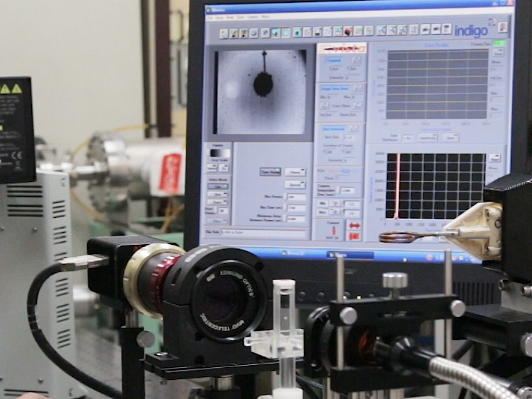
この稿では実験装置と誘導; を介して加熱小さな磁性球の NIR ベース温度イメージング技術の手順また、それは 2 つの代表的な 2 D の吸光度の画像の結果を示します。Λでキャプチャ 10.0 mm 厚さの水層に直径 2.0 mm の鋼球の 1 つのイメージは 1150 nm を =。2 番目の画像はλでキャプチャ 2.0 mm 厚マルトース シロップ層に直径 0.5 mm の鋼球 = 1412 nm。また述べる計算法と温度の三次元 (3 D) の半径方向分布の結果逆アベル変換 (IAT) を吸光度 2 D 画像に適用することによって。IAT は、3次元温度分布が球対称加熱球 (図 2)19の場合のようにすると見なされますときに有効です。IAT 計算のためマルチ ガウス関数をフィッティング法を採用、ここで解析的25,26,27,28,29 ガウス関数の Iat を取得することができますのでとデータを単調減少にもフィットこれは、単一の熱源からの熱伝導を用いた実験が含まれています。
Protocol
Representative Results
Discussion
本稿で紹介しているテクニックの水の近赤外吸収の温度依存性を使用して 1 つ新規であり、必要な機器と実装の設定で重大な困難を渡さない。入射光はハロゲン ランプと、NBPF を使用して簡単に製作することができます。ただし、レーザーを使用することはできません、コヒーレント干渉パターン画像に表示されますので。一般的な光学レンズおよび可視光用ガラス細胞使用できますが、 <em…
Divulgaciones
The authors have nothing to disclose.
Acknowledgements
著者は、山田健太、藤岡亮太氏、氏瑞希許田を実験・ データ解析に応援ありがちましょう。この作品は、日本学術振興会科研費助成番号 25630069、スズキ財団と精密測定技術振興財団、日本によって支えられました。
Materials
| Induction heating system | CEIA, Italy | SPW900/56 | 780 kHz, 5.6 kW (max). |
| Coil | SA-Japan | custom | Water-cooled copper tube; two-turn; outer dia. 28 mm. |
| Water chiller | Matsumoto Kikai, Japan | MP-401CT | |
| Halogen lamp | Hayashi Watch-Works, Japan | LA-150UE-A | |
| Narrow bandpass filter for λ = 1150 nm | Andover | 115FS10-25 | Full width at half-maximum (FWHM): 10 nm. |
| Narrow bandpass filter for λ = 1412 nm | Andover | semi-custom | Full width at half-maximum (FWHM): 10 nm. |
| Bandpass filter for λ = 850−1300 nm | Spectrogon | SP-1300 | |
| Bandpass filter for λ = 1100−2000 nm | Spectrogon | SP-2000 | |
| NIR camera | FLIR Systems | Alpha NIR | InGaAs |
| Image acquisition software | FLIR Systems | IRvista | |
| Image processing software | NIH | ImageJ | ver. 1.51r |
| Image processing software | MathWorks | Matlab | ver. 2016a |
| Telecentric lens | Edmond Optics | 55350-L | X1 |
| Steel sphere (0.5 mm dia.) | Kobe Steel, Japan | Fe-1.5Cr-1.0C-0.4Mn (wt %) | |
| Steel sphere (2.0 mm dia.) | Kobe Steel, Japan | Fe-1.5Cr-1.0C-0.4Mn (wt %) | |
| Maltose syrup as aqueous gel | Sonton, Japan | Mizuame | Food product |
Referencias
- Moros, E. G. . Physics of Thermal Therapy. , (2012).
- Périgo, E. G., Hemery, G., Sandre, O., Ortega, D., Garaio, E., Plazaola, F., Teran, F. J. Fundamentals and advances in magnetic hyperthermia. Appl Phys Rev. 2, 041302 (2015).
- Bardati, F., Marrocco, G., Tognolatti, P. Time-dependent microwave radiometry for the measurement of temperature in medical applications. IEEE Trans Microwave Theo Tech. 52, 1917-1924 (2004).
- Levick, A., Land, D., Hand, J. Validation of microwave radiometry for measuring the internal temperature profile of human tissue. Meas Sci Technol. 22, 065801 (2011).
- Daniels, M. J., Varghese, T., Madsen, E. L., Zagzebski, J. A. Non-invasive ultrasound-based temperature imaging for monitoring radiofrequency heating-phantom results. Phys Med Biol. 52, 4827 (2007).
- Daniels, M. J., Varghese, T. Dynamic frame selection for in vivo ultrasound temperature estimation during radiofrequency ablation. Phys Med Biol. 55, 4735 (2010).
- Seo, C. H., Shi, Y., Huang, S. -. W., Kim, K., O’Donnell, M. Thermal strain imaging: A review. Interface Focus. 1, 649-664 (2011).
- Bayat, M., Ballard, J. R., Ebbini, E. S. Ultrasound thermography: A new temperature reconstruction model and in vivo results. AIP Conf Proc. 1821, 060004 (2017).
- Petrova, E., Liopo, A., Nadvoretskiy, V., Ermilov, S. Imaging technique for real-time temperature monitoring during cryotherapy of lesions. J Biomed Opt. 21, 116007 (2016).
- Gardner, B., Matousek, P., Stone, N. Temperature spatially offset Raman spectroscopy (T-SORS): Subsurface chemically specific measurement of temperature in turbid media using anti-Stokes spatially offset Raman spectroscopy. Anal Chem. 88, 832-837 (2016).
- Yoshioka, Y., Oikawa, H., Ehara, S., Inoue, T., Ogawa, A., Kanbara, Y., Kubokawa, M. Noninvasive measurement of temperature and fractional dissociation of imidazole in human lower leg muscles using 1H-nuclear magnetic resonance spectroscopy. J Appl Physiol. 98, 282-287 (2004).
- Galiana, G., Branca, R. T., Jenista, E. R., Warren, W. S. Accurate temperature imaging based on intermolecular coherences in magnetic resonance. Science. 322, 421-424 (2008).
- Rapoport, E., Pleshivtseva, Y. . Optimal Control of Induction Heating Processes. , (2006).
- Lucía, O., Maussion, P., Dede, E. J., Burdío, J. M. Induction heating technology and its applications: Past developments, current technology, and future challenges. IEEE Trans Ind Electron. 61, 2509-2520 (2014).
- Kakuta, N., Kondo, K., Ozaki, A., Arimoto, H., Yamada, Y. Temperature imaging of sub-millimeter-thick water using a near-infrared camera. Int J Heat Mass Trans. 52, 4221-4228 (2009).
- Kakuta, N., Fukuhara, Y., Kondo, K., Arimoto, H., Yamada, Y. Temperature imaging of water in a microchannel using thermal sensitivity of near-infrared absorption. Lab Chip. 11, 3479-3486 (2011).
- Kakuta, N., Kondo, K., Arimoto, H., Yamada, Y. Reconstruction of cross-sectional temperature distributions of water around a thin heating wire by inverse Abel transform of near-infrared absorption images. Int J Heat Mass Trans. 77, 852-859 (2014).
- Kakuta, N., Yamashita, H., Kawashima, D., Kondo, K., Arimoto, H., Yamada, Y. Simultaneous imaging of temperature and concentration of ethanol-water mixtures in microchannel using near-infrared dual-wavelength absorption technique. Meas Sci Technol. 27, 115401 (2016).
- Kakuta, N., Nishijima, K., Kondo, K., Yamada, Y. Near-infrared measurement of water temperature near a 1-mm-diameter magnetic sphere and its heat generation rate under induction heating. J Appl Phys. 122, 044901 (2017).
- Libnau, F. O., Kvalheim, O. M., Christy, A. A., Toft, J. Spectra of water in the near- and mid-infrared region. Vib Spectrosc. 7, 243-254 (1994).
- Siesler, H. W., Ozaki, Y., Kawata, S., Heise, H. M. . Near-Infared Spectroscopy. , (2002).
- Shakher, C., Nirala, A. K. A review on refractive index and temperature profile measurements using laser-based interferometric techniques. Opt Laser Eng. 31, 455-491 (1999).
- Assebana, A., Lallemanda, M., Saulniera, J. -. B., Fominb, N., Lavinskaja, E., Merzkirchc, W., Vitkinc, D. Digital speckle photography and speckle tomography in heat transfer studies. Opt Laser Technol. 32, 583-592 (2000).
- Ambrosini, D., Paoletti, D., Spagnolo, S. G. Study of free-convective onset on a horizontal wire using speckle pattern interferometry. Int J Heat Mass Trans. 46, 4145-4155 (2003).
- Bracewell, R. N. . The Fourier Transform and Its Applications. , (2000).
- Yoder, L. M., Barker, J. R., Lorenz, K. T., Chandler, D. W. Ion imaging the recoil energy distribution following vibrational predissociation of triplet state pyrazine-Ar van der Waals clusters. Chem Phys Lett. 302, 602-608 (1999).
- De Colle, F., de Burgo, C., Raga, A. C. Diagnostics of inhomogeneous stellar jets: convolution effects and data reconstruction. Astron Astrophys. 485, 765-772 (2008).
- Green, K. M., Borrás, M. C., Woskov, P. P., Flores, G. J., Hadidi, K., Thomas, P. Electronic excitation temperature profiles in an air microwave plasma torch. IEEE Trans Plasma Sci. 29, 399-406 (2001).
- Bendinelli, O. Abel integral equation inversion and deconvolution by multi-Gaussian approximation. Astrophys J. 366, 599-604 (1991).
- Dorband, B., Muller, H., Gross, H., Gross, H. Vol. 5 Metrology of Optical Components and Systems. Handbook of Optical System. , (2012).
- Sheikholeslami, M., Rokni, H. B. Simulation of nanofluid heat transfer in presence of magnetic field: A review. Int J Heat Mass Trans. 115, 1203-1233 (2017).
- Häfeli, U., Schütt, W., Teller, J., Zborowski, M. . Scientific and Clinical Applications of Magnetic Carriers. , (2013).