Engineering Chimeric Antigen Receptor-Natural Killer Cells Targeting Fungal Infections Using the Non-viral Sleeping Beauty Transposon System
Summary
We outline a transfection protocol for producing chimeric antigen receptor-natural killer (CAR-NK) cells targeting fungal pathogens using the non-viral Sleeping Beauty transposon system. To assess antigen-specific activation, we co-cultured the engineered cells with Aspergillus fumigatus germ tubes and measured IFN-γ secretion.
Abstract
Chimeric antigen receptor (CAR)-based cell therapies have shown impressive efficacy in the treatment of hematological malignancies. Recently, these therapies are being developed for infectious diseases, yet studies targeting fungal infections remain scarce. To identify optimal targets and optimize cellular products, we developed a method to engineer chimeric antigen receptor-natural killer (CAR-NK) cells and evaluated their response to stimulation by fungi. This paper describes a straightforward and robust method for generating CAR-NK cells tailored to fungal targets using the non-viral Sleeping Beauty transposon system. NK-92 cells are transfected with the hyperactive transposase SB100X vectorized as minicircle DNA (MC) along with a plasmid-encoded CAR transposon. Transfection efficiency is assessed 1 week later using flow cytometric analysis. Prior to functional testing, the cells expressing the transgene are enriched using magnetic-activated cell sorting and cultured for 1 more week. To evaluate antigen-specific activation, the engineered cells are co-cultured with Aspergillus fumigatus germ tubes for at least 6 h. Subsequently, the concentration of the secreted interferon-gamma (IFN-γ) is measured using an enzyme-linked immunosorbent assay.
Introduction
Aspergillus fumigatus (A. fumigatus) is a ubiquitous fungus, with the average person inhaling between 100 and 1,000 conidia on a daily basis1. In patients with acquired or congenital immunodeficiencies, such as those undergoing chemotherapy or who are suffering from leukemia, A. fumigatus can result in severe invasive apsergillosis. Each year, more than 200,000 people worldwide contract aspergillosis. Despite the use of antimycotics for prevention and therapy with azole derivatives, the mortality rate remains between 30% and 95%2,3. Additionally, the treatment has significant side effects, and 7% of A. fumigatus cultures are already azole-resistant4.
Therapy with chimeric antigen receptor (CAR)-engineered T cells has shown clinical success in treating malignant diseases and achieved promising preclinical results in combating infectious diseases such as invasive aspergillosis5,6,7,8. Our previously described Af-CAR-T cells targeting a protein in the cell wall of A. fumigatus hyphae have shown promising results in preclinical models6.
The reduced risk of graft versus host disease, cytokine release syndrome, and neurotoxicity makes CAR-NK cells a valuable alternative to CAR-T cells as an "off-the-shelf" treatment for acute infections9. Additionally, NK cells have been shown to play a crucial role in the antifungal immune response. When stimulated with A. fumigatus, NK cells secrete cytokines such as IFN-ɣ and chemokines like CCL-3 and CCL-4 and release cytotoxic granules10,11.
The NK-92 cell line is a popular source of NK cells for genetic engineering, offering the advantage of rapid proliferation and expansion in relatively simple culture conditions12. Several preclinical studies have demonstrated the therapeutic potential of CAR-transduced NK-92 cells in treating both hematological and solid tumors. Numerous clinical trials are ongoing to investigate their safety, efficacy, and potential use as "off-the-shelf" cellular therapy13.
Achieving high transfection efficiencies and stable transgene expression in NK cells is challenging. While viral methods promise high efficiencies, they carry the risk of insertion mutations with subsequent oncogenesis9,14. The latest generation of Sleeping Beauty (SB) transposon vectors offers a safe, potent, and economically viable system for stable gene transfer, overcoming limitations of viral and transient non-viral gene delivery methods15.
Therefore, this protocol outlines a novel methodology for the generation of Af-CAR-NK-92 cells, which can potentially serve as an "off-the-shelf" therapeutic option for patients with life-threatening invasive aspergillosis. The efficacy of this approach is exemplified by the increased production of IFN-ɣ upon stimulation with A. fumigatus hyphae.
Protocol
All experiments described here must be conducted under sterile conditions. In this protocol, we show two conditions: (I) Mock transfection without DNA that will serve as control and (II) CAR transfection using the previously described Af-CAR6. The Af-CAR plasmid contains a truncated epidermal growth factor receptor (EGFRt) sequence downstream of the CAR, which will be used as a transfection marker (Supplemental Figure S1). For each condition, 4 × 10⁶ NK-92 cells are required.
1. Culturing the NK-92 cells
- One or two days before the transfection, prepare fresh cell culture medium with the following composition: 75% (v/v) alpha-MEM Medium (with nucleosides) + 12.5% (v/v) heat-inactivated fetal bovine serum (FBS) + 12.5% (v/v) heat-inactivated horse serum + 2 mM L-glutamine.
- Re-suspend at least 107 NK-92 cells at a cell density of 5 × 10⁵ cells/mL.
- Add IL-2 to the cell suspension at a concentration of 150 UI/mL.
- Culture the cells in a humidified incubator at 37 °C with 5% CO2.
NOTE: IL-2 can be used at a final concentration of 5 ng/mL.
2. Non-viral transfection
- Check the quality of the cells under the microscope. A suspension of cell clusters and a clear background indicate a healthy cell culture.
- In a 6-well plate, add 2 mL of cell culture medium per well for each condition. Place the plate in a humidified incubator at 37 °C with 5% CO2.
NOTE: Antibiotics can reduce cell viability after transfection. Keep the medium antibiotic-free. - Count the cells and check the cell viability using (e.g., Trypan Blue staining).
- Transfer 8 × 10⁶ NK-92 cells to a 15 mL conical bottom tube. Centrifuge at 200 × g for 5 min with an acceleration and deceleration of 3. Use this program for all the following centrifugation steps.
- During the centrifugation, prepare two 1.5 mL reaction tubes. Pipette the DNA for Af-CAR (8 µg of Plasmid-DNA and 4 µg of SB100X Mini Circle, Table 1) into one of the tubes. Keep the second one DNA-free to be a Mock control.
- When the centrifuge is ready, gently discard the supernatant and fill the tube containing the cells with up to 15 mL of prewarmed Phosphate Buffered Saline (PBS).
- Centrifuge (step 2.4).
- During the centrifugation, prepare one transfection system tube for each condition by adding 3 mL of electrolytic buffer in each tube. Put the first tube in the pipette station.
- When the centrifuge is ready, gently discard the supernatant and resuspend the cell pellet in 200 µL of resuspension buffer (100 µL/4 × 106 cells).
NOTE: Avoid leaving the cells in resuspension buffer for more than 15-30 min, as this lowers cell viability and transfection efficiency. - Add 100 µL of the cell suspension into each of the two reaction tubes. Mix gently.
NOTE: Avoid air bubbles during pipetting as air bubbles cause arcing, which leads to cell death. - Pipette the DNA-cell mixture from the first reaction tube with the transfection system pipette and insert the pipette vertically into the tube placed in the pipette station.
NOTE: Ensure that the tip fits properly. - Set the first pulse at 1,650 V and pulse time for 20 ms and press Start.
- After completion, set the second pulse at 500 V and pulse time for 100 ms. Press Start.
- After completion, slowly remove the pipette from the pipette station and immediately transfer the cells into one well of the prepared culture plate containing 2 mL of pre-warmed cell culture medium (see step 2.2). Gently move the plate circlewise to ensure that the cells are evenly distributed.
- For the Mock sample, take 100 µL of the cell suspension from the DNA-free reaction tube (see step 2.9) and repeat the same procedure from step 2.11 to step 2.14, using a new tube and a new pipette tip.
- Incubate the plate in a humidified incubator at 37 °C with 5% CO2.
- After 30 min of incubation, add IL-2 at a final concentration of 150 UI/mL.
3. Checking the transfection efficiency using flow cytometry
- After 1 week of cultivating and splitting the cells every second day, take 1-5 × 10⁵ cells of each condition into a polystyrene round-bottom tube.
- Wash the cells by adding 1 mL of buffer (PBS + 0.5% (v/v) FBS + 2 mM EDTA); centrifuge and discard the supernatant.
- Resuspend the cell pellet in 50 µL of buffer. Add 1 µL of 7-AAD to detect the dead cells.
- Add 5 µL of anti-tEGFR-Alexa fluor 647 antibody to stain the transfected cells.
- Incubate for 20 min at 4 °C.
- Wash the cells (see step 3.2).
- Resuspend the cell pellet in 200 µL of buffer.
- Measure the samples on a flow cytometer.
NOTE: If the transfection efficiency is not satisfying, proceed with the enrichment of transgene-expressing cells.
4. Enrichment of transgene-expressing cells by magnetic-activated cell sorting (MACS)
NOTE: For optimal results, start the protocol with 8-10 × 106 cells. All centrifugation steps are conducted at 4 °C.
- Prepare buffer as described in step 3.2. Dilute the cells at a concentration of 1-2 × 10⁶/100 µL with the buffer in a 15 mL conical bottom tube.
- Add 1 µL of anti-EGFRt-biotin per 1-2 × 10⁶ cells, and gently swirl the tube to ensure an evenly distributed solution.
- Incubate for 25 min at 4 °C, then fill the tube with up to 10 mL of buffer.
- Centrifuge. Discard the supernatant.
- Add 8 µL/1 × 106 cells of cold buffer.
- Add 2 µL of anti-biotin magnetic beads per 1 × 10⁶ cells.
- Incubate for 15 min at 4 °C.
- Wash by filling up to 10 mL with buffer.
- Centrifuge. Discard the supernatant and resuspend the cells in 500 µL of buffer.
- Place an LS column in the MACS magnet and wash with 3 mL of buffer.
- As soon as the buffer has completely migrated through the column, add the cell suspension.
- Wash the column 3x, each with 3 mL of buffer.
- Remove the column from the magnet and place it in a clean 15 mL conical bottom tube.
- Add 5 mL of buffer to the column. Using the plunger, flush out the EGFR-positive cells as quickly as possible.
- Culture the cells for a few days; then, check the efficiency by flow cytometry.
5. Functional testing
NOTE: For the upcoming experiment, prepare and use the following medium: RPMI + 20% FBS.
- Day 1 – Seeding Aspergillus fumigatus conidia
- Prepare an A. fumigatus conidia suspension at a concentration of 2.5 × 10⁵ conidium/mL in the medium.
- In a 96-well culture plate, pipette 100 µL of the conidia suspension per well and incubate at 25 °C for 16 h.
NOTE: After the incubation period, the conidia will germinate (Figure 1).
- Day 2 – Co-culture with CAR NK-92 cells
- Wash the Mock and Af-CAR NK-92 cells 2x with the medium.
- Co-culture with the fungus at an effector-to-target ratio of 2:1 by adding 5 × 10⁴ cells in 100 µL of medium per well (Figure 2).
- For positive control, add 1 ng of PMA and 100 ng of ionomycin per well in 100 µL of medium. Use cells in medium only as a negative control.
- Incubate the plate at 37 °C in a humidified CO2 incubator for at least 6 h.
- Centrifuge the plate at 300 × g for 5 min.
- Harvest 100 µL of each well; avoid touching the bottom. Transfer the supernatants to reaction tubes.
NOTE: Make sure that only the supernatant is pipetted and not the grown fungus. If in doubt, add another centrifugation step. - Perform IFN-ɣ ELISA following the manufacturer's instructions or store the samples at -80 °C until use.
Representative Results
The entire process of generating Af-CAR NK-92 cells, including transfection, recovery, enrichment, and expansion, takes approximately 14 days. After transfection, the cells are cultured and split every second day. Cell viability may decrease during the first splitting two days post transfection. Typically, the cells recover 4 days post transfection and begin proliferating, with a doubling time of 48 h.
Following recovery, transfection efficiencies are measured and are expected to reach 10-20% (representative results shown in Figure 3). Enrichment of EGFR-positive cells via MACS increases the percentage of Af-CAR NK-92 cells to over 95% (representative results shown in Figure 4).
Cultures with more than 95% Af-CAR cells were used for functional assays. Here, we co-cultured the cells with A. fumigatus germ tubes for 6 h and measured IFN-ɣ secretion as an activation marker. As shown in Figure 5, the Af-CAR cells exhibited significant cytokine secretion (mean = 340.6 ± 86.5 pg/mL) compared to Mock NK-92 cells (mean = 75.6 ± 19.5 pg/mL), indicating substantially higher activation of the Af-CAR cells (p-value = 0.03).
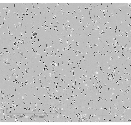
Figure 1: Germ tubes of Aspergillus fumigatus. Microscopy image of A. fumigatus after 16 h of incubation. Germ tubes/small hyphae are observed and distributed homogeneously around the well. Scale bar = 400 µm. Please click here to view a larger version of this figure.
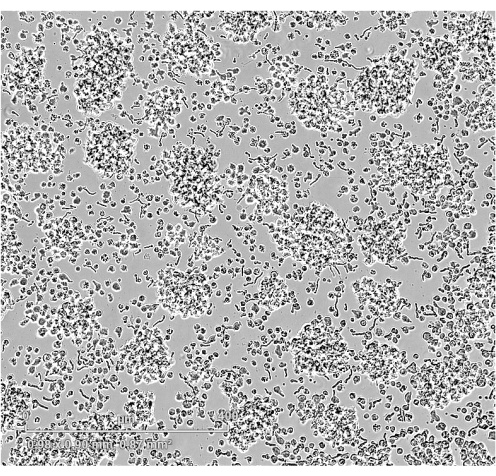
Figure 2: Co-culture of Aspergillus fumigatus germ tubes with NK-92 cells. Microscopy image of co-culture. The NK-92 cells form clusters and are in contact with the A. fumigatus germ tubes. Scale bar = 400 µm. Please click here to view a larger version of this figure.
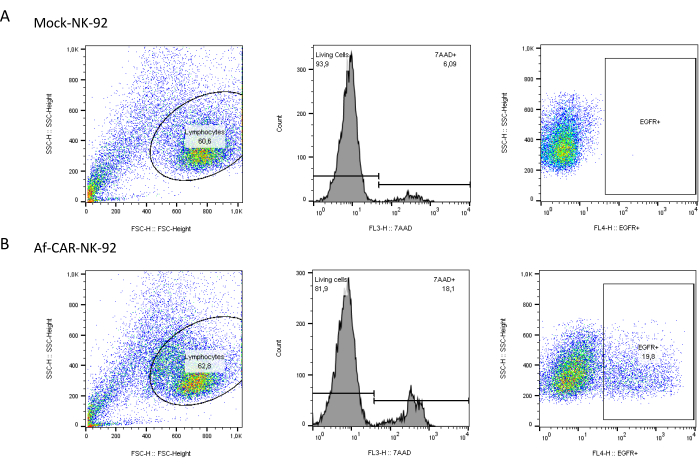
Figure 3: Transfection efficiency evaluated by staining for the transfection marker EGFR. On day 5, after electroporation, NK-92 cells were stained with 7-AAD and anti-EGFR. The results are shown for one representative experiment for (A) Mock and (B) Af-CAR NK-92 cells. The left column shows dot plots of cell size vs cell granularity; the gate is set on the NK-92 cells. The dead cells are outgated using 7-AAD labeling (second column). The percentage of living EGFR+ cells is shown in the third column. Abbreviations: EGFR = epidermal growth factor receptor; 7-AAD = 7-aminoactinomycin D; CAR = chimeric antigen receptor. Please click here to view a larger version of this figure.
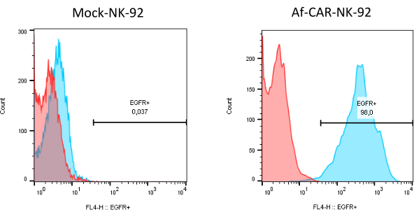
Figure 4: Cell surface staining results after enrichment of NK-92 cells expressing the transgene. On the left, the Mock cells are displayed. On the right, the Af-CAR-NK cells are shown. The red histograms represent the unstained cells. The blue histograms represent the corresponding stained cells. The gate is set on the EGFR+ cells; the numbers show the percentage of positive cells. Abbreviations: EGFR = epidermal growth factor receptor; CAR = chimeric antigen receptor. Please click here to view a larger version of this figure.
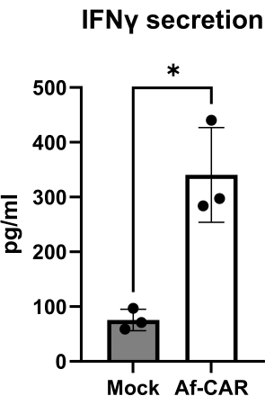
Figure 5: Cytokine secretion of NK cells activated by Aspergillus fumigatus germ tubes. The diagrams present results of IFN-ɣ secretion of Af-CAR and Mock cells in co-culture with fungus measured by ELISA after 6 h of co-culture. The data shown are the mean ± SD of three independently performed experiments (n = 3). P-value calculated by Welch's t-test using GraphPad Prism 10, * p < 0.05. Abbreviations: NK = natural killer; IFN-ɣ = interferon gamma; CAR = chimeric antigen receptor; ELISA = enzyme-linked immunosorbent assay. Please click here to view a larger version of this figure.
| Name | Company | Stock concentration | Reference | |
| Transposase | SB100X Mini Circle | PlasmidFactory | 1 µg/µL | 19 |
| Transposon | Af-CAR plasmid | Produced in house | 1 µg/µL | 6 |
Table 1: DNA used in this protocol.
Supplemental Figure S1: Plasmid map of the Af-CAR transposon: Schematic of plasmid vector and design of CAR transposon. Abbreviations: EF1 = elongation factor-1 alpha promotor; ORI = bacterial origin of replication; LIR = left inverted repeat; RIR: right inverted repeat; Cyto = Cytoplasmic; Tm = Transmembrane. Please click here to download this File.
Discussion
The provided protocol offers a simple and reliable method for creating CAR-NK cells that target fungal pathogens using the non-viral Sleeping Beauty transposon system. This method allows for the stable and permanent insertion of large transgenes, which enables the expansion of cells to obtain the necessary quantities for further analysis14,16.
Several key factors can impact the success of the transfection, including cell number and viability, pulse strength and duration, as well as DNA quality and quantity17. It is crucial to assess the cells' health before beginning, ensuring a viability of at least 90%. It has been demonstrated that an elevated pulse strength can enhance the transfection efficiency of a cell population. However, this approach can also result in a significant reduction in cell viability. In instances with excess cells and a limited quantity or quality of DNA, the number of cells that successfully take up the plasmid may be insufficient17. Nevertheless, high concentrations of DNA have been demonstrated to be toxic and reduce cell viability18.
Previous studies have achieved higher transfection efficiencies but focused solely on transient protein expression17. In contrast, our protocol ensures stable transgene insertion. To address low transfection efficiencies, we implemented the enrichment of positively transfected cells and achieved approximately 95% CAR-expressing cells. However, this augmented the production time to ~1 week. If shorter production times are desired, the transposon DNA could be delivered as a minicircle, which can enhance transfection efficiency and may eliminate the need for the enrichment step19. Nevertheless, the production time cannot be reduced to <1 week to allow for cell recovery and proliferation.
Given the acute nature of invasive aspergillosis, "off-the-shelf" cellular therapies would be beneficial. The challenges and costs associated with isolating cells from patients and expanding primary cells have increased the popularity of NK-92 as a convenient cell line for CAR-NK cell production20. NK-92 cells are generally robust and proliferate rapidly in the presence of IL-2, which facilitates the attainment of large cell quantities in a relatively short period. In light of these advantages, we have developed a protocol to gene-engineer NK-92 cells, demonstrating that it is possible to safely transfect and prepare CAR-NK cellular products with high purity. While this protocol could be adapted to prepare primary CAR-NK cells, several modifications should be considered. These include optimizing the number and intensity of pulses, the amount of DNA used, and establishing a suitable expansion protocol. Any modifications to the protocol would require a thorough investigation to ensure efficacy. Nevertheless, our protocol provides a foundational template that could be adapted for primary NK cells in future experiments.
In summary, this innovative transfection protocol for producing stable Af-CAR-NK cells is an efficient and reproducible method that can be employed to target other diseases. Additionally, it could be used as a cost-effective method to screen for novel CARs targeting a variety of antigens.
Disclosures
The authors have nothing to disclose.
Acknowledgements
Funding and support for the project were provided by Wilhelm Sander Stiftung, project 2020.017.1 to M.H., and J.L.; Deutsche Forschungsgemeinschaft (Collaborative Research Center/Transregio 124 Pathogenic fungi and their human host: Networks of interaction-FungiNet; project A8 to M.H. and H.E.).
Materials
| 15 ML conical bottom tubes | Greiner Bio-One | 188271 | PP, 17/120 MM, CELLSTAR, STERILE |
| 50 ML conical bottom tubes | Greiner Bio-One | 227270 | PP, 30/115 MM, CELLSTAR, STERILE |
| 6-and 96-Well- Cell culture plate | Corning Inc. LifeSciences | 83-3736/ 83-3474 | TC-treated Multiple Well Plates, Flat bottoms, Treated for optimal cell attachment, Sterilized by gamma irradiation, Nonpyrogenic |
| 7-AAD (7-Aminoactinomycin D) | BD Biosciences | 559925 | eady-to-use nucleic acid dye for the exclusion of nonviable cells in flow cytometric assays |
| Alexa Fluor 647 anti-human EGFR Antibody | Biolegend | 352918 | Clone AY13 |
| Anti-Biotin MicroBeads UltraPure | Miltenyi Biotec | 130-105-637 | UltraPure MicroBeads conjugated to monoclonal mouse anti-biotin antibodies |
| Aspergillus fumigatus | ATCC | 46645 | |
| Biosphere Filter Tips (20 and 100 μL) | Sarstedt | 70.3030.365 / 70.3030.275 | |
| Dulbecco′s Phosphate Buffered Saline (PBS) | Sigma-Aldrich | D8537 | Modified, without calcium chloride and magnesium chloride, liquid, sterile-filtered, suitable for cell culture, endotoxin, tested |
| ELISA MAX Deluxe Set Human IFN-γ | Biolegend | 430104 | |
| Ethylenediaminetetraacetic acid disodium salt solution (EDTA) | Sigma-Aldrich | E7889 | for molecular biology, 0.5 M in H2O, DNase, RNase, NICKase and protease, none detected, 0.2 μm filtered. |
| FACS clean | BD Biosciences | 340345 | |
| FACS Flow Sheath Fluid | BD Biosciences | 342003 | |
| FACS Rinse Solution | BD Bioscience | 340346 | |
| Fetal Bovine Serum (FBS) | Sigma-Aldrich | F7524 | sterile-filtered, suitable for cell culture, heat inactivated before use |
| Gibco, MEM α, nucleosides | Thermofisher Scientific | 22571020 | Phenol Red, Ribonucleosides, Deoxyribonucleosides, Sodium Bicarbonate |
| Horse serum | Thermo Fisher Scientific | 16050122 | Sterile-filtered, heat inactivated before use |
| Human IL-2 IS, research grade | Miltenyi Biotec | 130-097-742 | Research grade. The ED50 is ≤0.3 ng/mL, corresponding to an activity of ≥3 × 10⁶ IU/mg. |
| Ionomycin | Sigma-Aldrich | I0634-1MG | from Streptomyces conglobatus, used as positive control for cytokine secretion |
| L-Glutamine | Sigma-Aldrich | W368401 | For cell culture |
| MACS LS Column | Miltenyi Biotec | 130-042-401 | Columns and plungers, sterile packed |
| NK-92 | DSMZ | ACC 488 | |
| Neon Transfektionssystem 100 μL-Kit | Thermo Fisher Scientific | MPK10096 | Resuspension Buffer R, Electrolytic Buffer E2, 100 μL Neon Tips, Neon Electroporation Tubes |
| Phorbol-12-myristat-13-acetat (PMA) | Sigma-Aldrich | P1585-1MG | used as positive control for cytokine secretion |
| Polystyrene round-bottom tube, 5 mL | BD Bioscience | S-452400 | used for flow cytometry |
| Reaction tube, 1.5 ML | Greiner Bio-One | 7,26,90,001 | PP, transparent, cap attached, with injected graduation. Sterilized before use. |
| RPMI 1640 Medium, GlutaMAX | Thermo Fisher Scientific | 72400021 | GlutaMax I, Phenol Red, HEPES, Sterile-filtered |
| TipOne 1000 μL XL Graduated Filter Tip | StarLab | S1122-1730 | |
| Equipment | |||
| BD FACSCalibur | BD Bioscience | ||
| CO2 Incubator C60 | Labotect | ||
| Heraeus Multifuge 3SR | Thermo Scientific | ||
| HydroFLEX microplate washer | Tecan | ||
| NanoQuant Infinite M200 Pro | Tecan | ||
| Neon Transfection System | Invitrogen | ||
| Pipettes | Eppendorf | ||
| Quadro MACS Separator | Miltenyi Biotec |
References
- Fang, W., Latgé, J. -. P. Microbe profile: Aspergillus fumigatus: A saprotrophic and opportunistic fungal pathogen. Microbiology (Reading). 164 (8), 1009-1011 (2018).
- Brown, G. D., et al. Hidden killers: Human fungal infections. Sci Transl Med. 4 (165), 165rv113 (2012).
- Sun, K. S., Tsai, C. F., Chen, S. C., Huang, W. C. Clinical outcome and prognostic factors associated with invasive pulmonary aspergillosis: An 11-year follow-up report from Taiwan. PLoS One. 12 (10), e0186422 (2017).
- Meis, J. F., Chowdhary, A., Rhodes, J. L., Fisher, M. C., Verweij, P. E. Clinical implications of globally emerging azole resistance in Aspergillus fumigatus. Philos Trans R Soc Lond B Biol Sci. 371 (1709), 20150460 (2016).
- Seif, M., Einsele, H., Löffler, J. CAR T cells beyond cancer: Hope for immunomodulatory therapy of infectious diseases. Front Immunol. 10, 2711 (2019).
- Seif, M., et al. CAR T cells targeting Aspergillus fumigatus are effective at treating invasive pulmonary aspergillosis in preclinical models. Sci Transl Med. 14 (664), eabh1209 (2022).
- Kumaresan, P. R., et al. Bioengineering T cells to target carbohydrate to treat opportunistic fungal infection. Proc Natl Acad Sci U S A. 111 (29), 10660-10665 (2014).
- Kumaresan, P. R., et al. A novel lentiviral vector-based approach to generate chimeric antigen receptor T cells targeting Aspergillus fumigatus. mBio. 15 (4), e0341323 (2024).
- Lu, H., Zhao, X., Li, Z., Hu, Y., Wang, H. From CAR-T cells to CAR-NK cells: A developing immunotherapy method for hematological malignancies. Front Oncol. 11, 720501 (2021).
- Robertson, M. J., Ritz, J. Biology and clinical relevance of human natural killer cells. Blood. 76 (12), 2421-2438 (1990).
- Ziegler, S., et al. CD56 is a pathogen recognition receptor on human natural killer cells. Sci Rep. 7 (1), 6138 (2017).
- Esmaeilzadeh, A., Hadiloo, K., Jabbari, M., Elahi, R. Current progress of chimeric antigen receptor (car) t versus car nk cell for immunotherapy of solid tumors. Life Sci. 337, 122381 (2024).
- Zhang, J., Zheng, H., Diao, Y. Natural killer cells and current applications of chimeric antigen receptor-modified NK-92 cells in tumor immunotherapy. Int J Mol Sci. 20 (2), 317 (2019).
- Hu, Y., Tian, Z. G., Zhang, C. Chimeric antigen receptor (CAR)-transduced natural killer cells in tumor immunotherapy. Acta Pharmacol Sin. 39 (2), 167-176 (2018).
- Hudecek, M., et al. Going non-viral: The Sleeping Beauty transposon system breaks on through to the clinical side. Crit Rev Biochem Mol Biol. 52 (4), 355-380 (2017).
- Amberger, M., Ivics, Z. Latest advances for the Sleeping Beauty transposon system: 23 years of insomnia but prettier than ever. BioEssays. 42 (11), 2000136 (2020).
- Ingegnere, T., et al. Human car nk cells: A new non-viral method allowing high efficient transfection and strong tumor cell killing. Front Immunol. 10, 957 (2019).
- Stacey, K. J., Ross, I. L., Hume, D. A. Electroporation and DNA-dependent cell death in murine macrophages. Immunol Cell Biol. 71 (Pt 2), 75-85 (1993).
- Monjezi, R., et al. Enhanced CAR T-cell engineering using non-viral Sleeping Beauty transposition from minicircle vectors. Leukemia. 31 (1), 186-194 (2017).
- Moscarelli, J., Zahavi, D., Maynard, R., Weiner, L. M. The next generation of cellular immunotherapy: Chimeric antigen receptor-natural killer cells. Transplant Cell Ther. 28 (10), 650-656 (2022).

.
