Automatically Generated
A Label-free Xenograft Model for Investigating the Behavior of Human Stem Cell Spheroids in Chick Embryos
Summary
Here, we describe a method to transplant and identify human cell spheroids into chick embryos. This xenograft model uses the embryonic microenvironment as a source of instructive signals to assay cell migration, differentiation, and tropism and is especially suited for the study of primary and/or heterogeneous cell populations.
Abstract
Xenografts are valuable methods to investigate the behavior of human cells in vivo. In particular, the embryonic environment provides cues for cell migration, differentiation, and morphogenesis, with unique instructive signals and germ layer identity that are often absent from adult xenograft models. In addition, embryonic models cannot discriminate self versus non-self tissues, eliminating the risk of rejection of the graft and the need for immune suppression of the host. This paper presents a methodology for transplantation of spheroids of human cells into chicken embryos, which are accessible, amenable to manipulation, and develop at 37 °C.
Spheroids allow the selection of a specific region of the embryo for transplantation. After being grafted, the cells become integrated into the host tissue, allowing the follow-up of their migration, growth, and differentiation. This model is flexible enough to allow the utilization of different adherent populations, including heterogeneous primary cell populations and cancer cells. To circumvent the need for prior cell labeling, a protocol for the identification of donor cells through hybridization of human-specific Alu probes is also described, which is particularly important when investigating heterogeneous cell populations. Furthermore, DNA probes can be easily adapted to identify other donor species. This protocol will describe the general methods for preparing spheroids, grafting into chicken embryos, fixing and processing tissue for paraffin sectioning, and finally identifying the human cells using DNA in situ hybridization. Suggested controls, examples of interpretation of results and various cell behaviors that can be assayed will be discussed in addition to the limitations of this method.
Introduction
Xenografts are useful tools to investigate the behavior of human cells in vivo. These models have provided invaluable information for a wide range of scientific topics, such as the biology of human stem cells1, the observation of cellular events in real time2, and the investigation of tumoral angiogenesis and metastasis3. In addition, several aspects of cancer biology, including the tumorigenesis of patient-specific xenografts, have been studied4,5. Each of these xenograft models has their advantages and disadvantages and, thus, each one is better suited for specific scientific questions. Chick embryos are a popular developmental biology model as they are an accessible amniote model that is amenable to surgical manipulation. Heterologous grafts have allowed researchers to create precise fate maps6 or explore whether a trait is cell-autonomous or instructed by the environment7,8. A similar rationale allows the chick embryo to be used as a xenograft model to study the behavior of human cells.
The embryonic environment actively orchestrates tissue morphogenesis with migration and differentiation signals, as well as cell-cell interactions. Thus, compared to adult xenograft models, the embryo provides a more instructive milieu to assay the behavior of grafted cells, for example, by mimicking signals present in adult stem cell niches (e.g., BMPs, WNTs, NOTCH, and SHH9). In addition, the absence of an adaptive immune system during early development allows xenografts to be performed without the risk of an immune response or rejection of the donor tissue10. Previous studies have investigated xenografts of human cells into chicken embryos for this purpose. The neurogenic potential of human stem cells has been assayed after injection into the neural tube or blood vessels11 in addition to the integration of embryonic stem cells12 and induced pluripotent stem cells13 into the embryo. Human melanoma cells have also been studied using the chick's embryonic environment, which revealed links between their tumorigenesis and the behavior of neural crest cells14, as well as the reprogramming of the tumor cells with the information from the embryo15. This paper describes a protocol that is especially suited for studying the behavior of human primary and heterogeneous cell populations.
In the last decades, the stromal component of diverse tissues has been studied as an autologous source of progenitor/stem cells and for its proangiogenic and immunoregulatory properties, previously known as "mesenchymal stem cells"16,17,18. The first of these cell populations to be characterized was the bone marrow stromal/stem cell population (BMSCs), which have osteo-, adipo- and, to a lesser extent, chondrogenic potential in vivo19,20. Adipose-derived stromal cells (ADSCs) are a heterogeneous population obtained by enzymatic digestion of the lipoaspirate or dermolipectomy samples, followed by isolation of the stromal-vascular fraction (SVF) and finally expansion in culture21. In culture, these cells are phenotypically characterized by markers shared with other mesenchymal populations, such as CD90, CD73, CD105, and CD44, unique markers such as CD36, and the absence of hematopoietic (CD45) or endothelial (CD31) markers22. Additionally, ADSCs have osteo-, adipo-, and chondrogenic potential in vitro, and the number of stem/progenitor cells in this population can be defined by the fibroblastoid colony-forming unit (CFU-F) assay22. In vivo, cells with the ADSC phenotype have been reported to exist in stromal23 and/or perivascular24 compartments. It is becoming increasingly clear that, despite sharing markers after in vitro culture, the stromal compartment of different tissues reflects intrinsic characteristics of a given organ, and these cell populations have distinct properties depending on their source17,25,26,27. Furthermore, as these cells are isolated based on their adhesion to a cell culture dish, they may be composed of cells from diverse germ layers28. Thus, employing a xenograft method to study the differentiation potential and tropism of stromal cells in an unbiased way can provide valuable information about these cell populations to guide the development of future cell therapies.
The protocol described here (Figure 1) is a xenograft method that takes advantage of the low cost and ease of manipulation of chick embryos. It has been previously used to study the behavior of human ADSC29, skin fibroblasts29, menstrual blood-derived stromal cells30, and glioblastoma cells31. This method will include the transplantation of cells as spheroids32, which can be prepared from any population of adherent cells (Figure 2). Surgical procedures and the preparation of custom surgical materials-the microscalpels and glass capillaries-will also be described (Figure 3). Human cells are detected in histological sections by hybridizing human-specific Alu probes (Figure 4), thus eliminating the need for prior labeling of the grafted cells. The representative results describe the behavior of human ADSC grafted both in the somitic region at the wing bud level (Figure 5, Figure 6, and Figure 7) and the first pharyngeal arch (Figure 8), as well as human primary glioblastoma spheroids grafted in the prosencephalon (Figure 8). Cell migration, differentiation, and interaction with chick embryonic tissues will be described, as well as suggested assays to further investigate cell behavior using co-staining or staining of adjacent sections.
Protocol
All in vivo procedures used in this study complied with all relevant experimental guidelines for animal testing and research, in accordance with the Brazilian experimental animal use guidelines (L11794). The protocols used for handling chicken embryos were all approved by the Ethics Committee on the Use of Animals in Scientific Experimentation (Health Sciences Centre of the Federal University of Rio de Janeiro). The use of human cells was approved by the Ethics Committee of the University Hospital Clementino Fraga Filho (numbers 043/09 and 088/04). Specific pathogen-free (SPF) eggs of White Leghorn chicken (Gallus gallus) were used.
1. Preparation of cell spheroids
NOTE: Cell spheroids can be prepared with a wide range of cell types as long as they are adherent. For this protocol, human adipose-derived stromal cells (ADSCs) were isolated as previously described21,27 will be used (Figure 2A). Cells were obtained by digesting adipose tissue fragments or lipoaspirates with collagenase IA for 1 h at 37 °C under agitation, followed by plating at 1−2 × 10⁴ cells/cm2 and overnight incubation. Non-adherent cells were discarded, and the adherent cells were expanded for 3-6 passages. ADSCs were homogeneous for the expression of the surface antigens CD105, CD90, CD13, and CD44 and negative for hematopoietic antigens CD45, CD14, CD34, CD3, and CD1927. While the method described here can be performed easily with minimal materials beyond what is routinely used for cell culture, optimal aggregation time and the need for partial dissociation should be determined empirically. Alternative methods for preparing cell spheroids may be employed, such as the hanging-drop method33 or agarose-coated wells34; see the discussion for more details. All procedures should be performed on a clean bench employing aseptic techniques.
- Warm up sterile PBS (phosphate-buffered saline), culture medium, and trypsin solution to 37 °C before cell manipulation.
- Culture ADSCs in low glucose DMEM supplemented with 10% fetal bovine serum (FBS), 2 mM L-glutamine, 100 U/mL penicillin, and 100 µg/mL streptomycin. Use a confluent 25 cm2 flask of ADSCs for the preparation of two plates of spheroids. Discard the culture medium and wash the culture dish three times with an appropriate amount of sterile PBS (5 mL PBS for each wash if a 25 cm2 cell culture flask is used).
- Add enough trypsin solution (containing 0.78 mM EDTA) to cover the cells (1 mL of trypsin solution if a 25 cm2 cell culture flask is used) and let the culture flask sit for 5 min at room temperature.
- Gently pipette the cells up and down to dissociate them. Add the same amount of culture medium containing FBS to inactivate the trypsin, and transfer the cell suspension to a 15 mL conical tube.
- Centrifuge for 10 min at 180 × g. Discard the supernatant, and tap the tube to loosen the pellet.
- Resuspend the cells in 500 µL-2 mL of culture medium adequate for the cell type (see step 1.2).
NOTE: The minimum concentration is 5 × 10⁵ cells/mL for adherent cells such as ADSCs and fibroblasts. A confluent 25 cm2 flask of ADSCs should be resuspended in 2 mL of medium and plated in 2 Petri dishes, using 1 mL each. Less adherent cells, such as glia, may benefit from plating at ~2 × 10⁶ cells/mL in a lower volume. - Carefully transfer the cell suspension to one side of a sterile 60 mm Petri dish (uncoated and untreated, such as those used for preparing agar plates). Try to restrict the culture medium to the smallest possible area (Figure 1B) by propping the dish over a piece of folded, clean gauze to keep it tilted in the incubator.
- Incubate the cell suspension at 37 °C, 5% CO2, until cell aggregates are formed (6-8 h after plating the ADSCs).
NOTE: Spheroid aggregation time may vary according to cell type. - If required (see discussion), partially dissociate large cell aggregates by gently pipetting up and down using 1000 µL tips. Dissociate ADSC spheroids 6-8 h after plating.
- Place the plate in the incubator until the spheroids are ready to be transplanted: round with defined edges and not easily dispersed (Figure 1C,D).
NOTE: ADSC spheroids are ready 2 days after partial dissociation.
2. Transplantation of spheroids into chick embryos
- Preparation
- Prepare chicken eggs by incubating 15-30 eggs in a humid incubator at 37.5 °C until the desired Hamburger-Hamilton (HH) stage is reached35: 40-45 h for grafts in somites at the limb bud level (HH11-12 or 13-19 somites) or 29-33 h for grafts at the cephalic region (HH8-9 or 5-8 somites). Position the eggs horizontally, and draw a line with a pencil on the top of the eggs before incubation (Figure 3D).
NOTE: Incubation times may vary slightly according to individual incubators. Drawing a line will ensure that the top portion of the egg, where the embryo is found, can be easily identified if the egg is rotated before the experiment. - Prepare sharp needles ("microscalpels") by attaching a sewing needle to a metal needle holder (Figure 3B). Using an oiled whetstone, sharpen the needle until it resembles a small scalpel with a thin edge (Figure 3C). Alternatively, prepare a sharp tungsten needle by electrolysis36.
- Prepare glass capillaries using the flame of a Bunsen burner to briefly melt the thin portion of a glass Pasteur pipette. Remove the molten portion from the flames and stretch it to form a thin capillary. Carefully divide the capillary into two parts by breaking it with microforceps. Prepare a thin capillary for India ink injection and a thick capillary for transferring spheroids (Figure 3B).
NOTE: By varying the pulling strength, it is possible to create different capillary gauges. Capillaries should be prepared just before the surgery. - Prepare the working surface and other materials for egg manipulation (Figure 3A). Sterilize all surgical materials in a 100 °C oven overnight or with 70% ethanol immediately before use. Warm sterile PBS to 37 °C. Attach a 200 µL tip (with barrier) to the aspirator tube assembly for India ink injection and transferring spheroids.
NOTE: Use tips with barriers when transferring spheroids of human cells. - Just before the experiment, add 2 drops of India ink stock solution to the 30 mm Petri dish and mix it with 1 mL of sterile PBS. Remove the spheroid plate from the incubator and place it over an icebox for the rest of the experiment.
- Prepare chicken eggs by incubating 15-30 eggs in a humid incubator at 37.5 °C until the desired Hamburger-Hamilton (HH) stage is reached35: 40-45 h for grafts in somites at the limb bud level (HH11-12 or 13-19 somites) or 29-33 h for grafts at the cephalic region (HH8-9 or 5-8 somites). Position the eggs horizontally, and draw a line with a pencil on the top of the eggs before incubation (Figure 3D).
- Preparation of each egg
- Remove one egg from the incubator and place it over an egg holder. Make a small hole in the sharp end of the egg (Figure 2D) and aspirate 1.5 mL of albumen using a syringe and needle. Insert the needle perpendicular to the egg to avoid damaging the egg yolk. Seal the hole using adhesive tape. Optionally, repeat this step for all eggs before the experiment, and reincubate the remainder of the eggs.
- Carefully cut open a window on the top of the egg using scissors (Figure 3D). Using the pipette, add 1-2 drops of PBS over the yolk. If necessary, remove large albumen bubbles (such as the ones seen in Figure 3F) using the pipette.
- Attach the thin capillary to the aspirator tube assembly. Fill the glass capillary partially with the India ink solution by aspiration. Inject enough India ink into the yolk under the embryo until the embryonic structures are seen (Figure 3E,F).
- Using the stereomicroscope, count the number of somites (Figure 4A). Individually number the eggs using a pencil (Figure 4B).
- Spheroid transplantation
- Identify the region of the graft: paraxial mesoderm at the wing bud level (presomitic mesoderm of the presumptive 15th to 20th somites37) (Figure 3G and Figure 5A) or presumptive first pharyngeal arch region (between the ectoderm and endoderm lateral to the posterior mesencephalon/first rhombomere38) (Figure 8A).
- Cut the vitelline membrane over the target region using a microscalpel.
- Using a pair of microscalpels, make a shallow cut in the region where the spheroid will be implanted (Figure 3H).
NOTE: Avoid damaging the underlying endoderm; otherwise, the yolk will leak (if this happens, discard the egg). - Remove the thin capillary from the aspirator and replace it with the thick capillary. Observe the spheroids under the stereomicroscope and choose a spheroid approximately the same size as the somite (Figure 2D). Aspirate this spheroid into the capillary.
- Gently deposit the spheroid next to the cut region (Figure 3H). Using the sharp needles, push the spheroid into the cut region (Figure 3I,J).
- Add 1-2 drops of PBS over the embryo using the pipette to ensure that the spheroid is firmly inserted.
NOTE: If the graft is dislodged, a new spheroid should be transplanted (steps 2.3.4 and 2.3.5). - Clean any albumin leak from the eggshell using tissue paper to ensure that the egg can be completely sealed, avoiding contamination. Seal the window in the egg using adhesive tape.
- Take note of the grafted region (Figure 4A). Carefully return the egg to the humid incubator at 37.5 °C to avoid dislodging the inserted spheroid.
- Incubate the egg until the desired stage.
NOTE: When the xenograft is performed into somites at the limb bud level, migration and cell death have been successfully assayed in 3.5-day-old embryos (HH21), and cell differentiation and tropism have been observed in 6-day-old embryos (HH29) (Figure 5A). Integration into host tissues has been performed in 8-day-old embryos as well (HH33). Donor cells grafted to the pharyngeal arch territory have been studied in 4.5-day-old embryos (HH25) (Figure 8A).
3. Tissue dissection, fixation, processing, and preparation of histological sections
- Dissection and fixation
- Prepare the fixative (at least 1 mL/embryo): ethanol 100%-formaldehyde 37%-glacial acetic acid in a 6:3:1 ratio. Clean the work surface, a pair of microforceps, surgical scissors or iris scissors, and a slotted spoon. Fill a 60 mm glass Petri dish with cold PBS and place it under a stereomicroscope.
- Open the egg by cutting out the adhesive tape.
- Cut the membranes around the embryo and remove it using the slotted spoon.
- Transfer the embryo to the Petri dish. If the embryo is 5-day-old (HH26) or older, promptly decapitate it before any manipulation ex ovo.
- Remove any remaining membranes using microforceps and scissors. Gently agitate the specimen in PBS to wash away any remaining yolk droplets.
- If the xenograft was performed at the wing level, discard the head. If cells were grafted to the cephalic region, cut and discard the lower half of the body, ensuring that the cardiac region is kept intact.
- Transfer each specimen to a separate 2.0 mL tube containing 1 mL of the fixative. Identify the tubes individually as before (Figure 4B). Incubate them overnight at 4 °C with agitation.
- Use a pellet of cells or spheroids (Figure 4F) as a positive control. For this, centrifuge a cell suspension (step 1.5) or cell spheroids (step 1.10) at 180 × g for 10 min in a 1.5 mL tube, discard the culture medium, and add 1 mL of the fixative without disturbing the pellet. Incubate the tube overnight at 4 °C without agitation, and proceed in the same manner as for the chick samples.
- Dehydration and embedding
- Dehydrate the embryos by successive washes of 1 mL of 70%, 80%, 90%, and 100% ethanol in 1x PBS for at least 1 h each with agitation.
- Incubate the samples overnight in 1 mL of 100% ethanol at room temperature with agitation.
- Incubate the samples in 1 mL of 100% xylene for 1 h in a fume hood. Repeat two more times.
- Transfer the contents of the tube to the staining block and discard the xylene. Add enough molten paraffin to cover the embryo and cover the staining block with its glass lid. Incubate overnight in a 65 °C oven.
- Prepare a warm plate and a mold for paraffin blocks for embedding.
NOTE: A pair of stretched-out paper clips are useful for positioning the embryo. - Fill the mold with molten paraffin and transfer the embryo to it. Position the embryo to obtain a transverse section of the trunk (Figure 5A) or a coronal section of the head (Figure 8A).
- Insert a paper name tag in the paraffin, opposite the surface to be sectioned, to help cut the embryo in the correct orientation (Figure 4C).
- If being used, place the embedding cassette over the sample. Let the block cool down completely before removing it from the mold. Alternatively, attach the hardened paraffin block to the cassette or other block holder using molten paraffin and let it cool down again.
- Sectioning
- Prepare the materials for sectioning. Warm up a hot plate to 42 °C and cover a cardboard or Styrofoam plate with clean aluminum foil approximately 30 cm x 20 cm. Clean all surfaces before use.
NOTE: Use gloves to avoid contamination with nucleases, especially if some sections will be used for RNA in situ hybridization. Avoid using a histology bath to stretch the paraffin sections for the same reason unless the water and surfaces can be thoroughly cleaned beforehand. - Trim the excess paraffin with a microtome blade. Create a bevel on both sides, in a trapezoidal shape, for easy separation of each section using a scalpel blade in a later step (Figure 3A).
- Attach the block to the microtome. Position the block carefully to ensure that the left and right sides are parallel to each other. Perform a finer adjustment after cutting a few sections of the sample and observing them under a microscope.
- When the target region has been reached (either the limb bud or first pharyngeal arch), cut 7 µm sections and place the paraffin ribbons over the plate covered with aluminum foil. Section the whole target region (Figure 4C).
- For preparing serial sections, cover the adequate number of slide glasses with drops of sterile deionized water. Transfer individual sections to the slides sequentially, using brushes and a scalpel. Transfer adjacent sections to different slides to create serial sections (Figure 4D). Prepare series of 3 slides for 3.5-day-old, 4 slides for 4.5-day-old, and 5 slides for 6-day-old embryos. Stain adjacent sections with different probes, antibodies, or classical histological stains.
NOTE: The same series may contain multiple adjacent slides if a large portion of the embryo is sectioned. Multiple sections can be transferred to each slide, keeping some space between sections to allow them to stretch. Do not place the slide on the warm plate yet. - After all sections have been transferred to the slides, add more water until all the sections are floating over a single water drop. Place the slide on a warm plate and let the sections stretch. Remove the slide from the warm plate, and remove the water by tilting the slide glass carefully against tissue paper.
NOTE: Do not let the sections directly touch the glass slide before they stretch. - Let the slides dry overnight in a 37 °C incubator.
- Prepare the materials for sectioning. Warm up a hot plate to 42 °C and cover a cardboard or Styrofoam plate with clean aluminum foil approximately 30 cm x 20 cm. Clean all surfaces before use.
4. Synthesis of digoxigenin-labeled Alu probes by polymerase chain reaction (PCR)
- Prepare a stock solution of the following human-specific primers39: AluFw: 5'-CGA GGC GGG TGG ATC ATG AGG T-3' and AluRev: 5'-TTT TTT GAG ACG GAG TCT CGC-3'.
- Prepare 50 µL of the following PCR reaction40: 1x PCR buffer, 2.0 mM MgCl2, 0.1 mM dCTP, 0.1 mM dGTP, 0.1 mM dATP, 0.065 mM dTTP, 0.035 mM dig-11-dUTP, 0.4 µM AluFw primers, 0.4 µM AluRev primers, 0.05 U/µL Taq polymerase, and 1 ng/µL human genomic DNA.
- Run the PCR using the following settings: initial denaturation at 94 °C for 4 min followed by 40 cycles of 94 °C for 20 s, 60 °C for 20 s, and 72 °C for 20 s, followed by a final denaturation of 72 °C for 5 min.
- Measure the probe concentration using a spectrophotometer. Store the probes at -20 °C.
NOTE: The PCR product should be 200-300 bp long after electrophoresis in a 2% agarose gel29.
5. Section in situ hybridization with Alu probes
- Day 1: Permeabilization and hybridization
NOTE: A sterilized slide glass jar should be used for all steps performed on day 1.- Preheat one wash of PBT (0.1% Tween 20 in PBS) in a 37 °C water bath and prepare a moist chamber with 50% formamide/50% deionized water.
NOTE: Calculate the volume of all washes based on the glass jar size. Unless specified, all washes are performed by immersion of the slides in the solution. - Remove the paraffin by three successive washes in xylene, 5 min each, in a fume hood.
- Rehydrate the series by two washes in 100% ethanol for 5 min each, followed by 90/70/30% ethanol washes in PBS for 2 min each.
- Wash in PBT (0.1% Tween 20 in PBS) three times for 5 min each.
- For permeabilization, add 2 µg/mL Proteinase K to the preheated PBT, immerse the slides in the solution, and incubate them for 14 min in a 37 °C water bath.
- Fix the sections by immersion in 4% paraformaldehyde/PBS for 20 min at room temperature.
- Wash with PBS for 5 min.
- After wiping excess PBS from each slide, cover the sections with 300 µL of hybridization buffer (50% deionized formamide, 4x SSC pH 5.0, 1x Denhardt's solution, 5% dextran sulfate, 100 µg/mL salmon sperm DNA) (Table 1). Incubate for 1 h at 42 °C in the formamide chamber.
- Prepare a solution of 0.2 ng/mL Alu probe in hybridization buffer. Tip the slide to remove the hybridization buffer and add 120 µL of the Alu probe solution over the sections. Cover with a glass coverslip, taking care to avoid bubbles.
NOTE: Parafilm should not be used to cover the slides in this step, as it will melt at temperatures above 70 °C. - Heat the slides on a hot plate at 95 °C for 5 min.
NOTE: Do not breathe the formamide fumes. - Incubate the slides at 42 °C in the formamide chamber overnight.
- Preheat one wash of PBT (0.1% Tween 20 in PBS) in a 37 °C water bath and prepare a moist chamber with 50% formamide/50% deionized water.
- Day 2: Stringency washes and immunohistochemistry
- Prepare 20x SSC buffer (3 M NaCl, 0.3 M sodium citrate, pH 7.5) (Table 1). Preheat two washes of 0.1x saline sodium citrate (SSC) buffer, pH 7.5, to 42 °C.
- Fill a slide glass jar with 2x SSC buffer, pH 7.5. Gently place the slides in the solution and wait for the coverslips to detach themselves. Remove the coverslips using pincers and incubate the slides in 2x SSC buffer for 5 min at room temperature.
- Rewash the slides with 2x SSC buffer, pH 7.5, for 5 min at room temperature.
- Wash the slides two times with 0.1x SSC buffer, pH 7.5, at 42 °C for 5 min each.
- Wash the slides two times with MABT (maleic acid buffer with Tween; 0.1 M maleic acid, 0.15 M sodium chloride, 0.1% Tween 20, pH 7.5) (Table 1) for 30 min each at room temperature.
- Wipe excess buffer from each slide and cover the sections with 400 µL of blocking solution (10% inactivated normal goat serum, 2% blocking reagent in MABT). Incubate for 2 h in a humid chamber (prepared with deionized water) at room temperature.
- Tip the slides to remove the liquid and add 150 µL of Fab anti-DIG fragments conjugated to alkaline phosphatase at a 1:2,000 dilution in blocking solution. Cover with a glass coverslip or parafilm and incubate in the humid chamber at 4 °C for 16 h.
- Day 3: Color development
- Gently remove the coverslips or parafilm inside a jar with MABT (as in step 5.2.2), and incubate the slides in MABT for 30 min at room temperature.
- Wash with MABT three more times for 30 min each.
- Wash with MAB (maleic acid buffer; 0.1 M maleic acid, 0.15 M sodium chloride, pH 7.5) (Table 1) for 30 min.
- Wash two times with NTM (NaCl-Tris-MgCl2 buffer; 100 mM Tris-HCl pH 9.5, 100 mM sodium chloride, 50 mM magnesium chloride) (Table 1) for 10 min each.
- Add the staining solution (0.45 µL/mL 4-Nitro blue tetrazolium chloride, 3.5 µL/mL 5-bromo-4-chloro-3-indolyl phosphate p-toluidine in NTM) (Table 1) to the staining jar and immerse the slides in the solution. Cover the jar with aluminum foil and let the color develop overnight at 37 °C.
- Day 4: Counterstaining and mounting
- Wash the slides three times with PBS at room temperature for 10 min each.
NOTE: Ensure that the staining solution is disposed of properly as halogen waste. - Mount the slides as they are by adding drops of an aqueous mounting media and covering the sections with a glass coverslip. Alternatively, proceed to either nuclear fast red or Alcian blue counterstaining.
- Wipe away excess PBS and add 500 µL of a nuclear counterstaining solution of 0.1% nuclear fast red41 (Table 1) over the sections and incubate the slides in a humid chamber for 10 min. Tip to remove excess nuclear fast red and proceed to dehydration (step 5.4.5).
- Immerse the sections in 0.5% Alcian blue solution (cartilage counterstain)42 for 10 min; rinse in distilled water and incubate in 1% phosphomolybdic acid for 10 min. Rinse with water again and proceed to dehydration.
NOTE: If the phosphomolybdic acid wash is omitted, Alcian blue becomes a nuclear counterstain. - Dehydrate in 70/95/100/100% ethanol for 5 min each.
- Wash with xylene three times for 5 min each time in a fume hood.
- Mount with a xylene-based mounting medium and glass coverslips.
- Let the slides dry for at least 16 h in a fume hood.
- Wash the slides three times with PBS at room temperature for 10 min each.
6. Image acquisition
- After the mounted slides are thoroughly dried, examine all sections for the presence of grafted human cells using an upright brightfield microscope (Figure 4E). Look for blue-purple Alu-positive cells (Figure 4F,G) and mark the sections containing Alu-positive cells using a marker pen (Figure 4E).
NOTE: Marking the sections with Alu-positive cells will make it easier to find them while photographing them and comparing adjacent slides stained with other markers. - Take care to photograph sections containing the xenograft in the same order as their placement on the slide (antero-posterior direction). Name each photo with all relevant information, such as [date of the xenograft]_[cell type]_[embryonic age]_[probe type]_[section number]_001. Include metadata files to add the scale bar later.
- If other slides from the same series are stained with another marker, e.g., RNA in situ hybridization or immunohistochemistry, photograph and name adjacent sections (Figure 6) in the same manner.
Representative Results
Identification of Alu-positive ADSCs in histological sections
Alu sequences are repetitive elements that comprise ~10% of the human genome and thus are excellent targets for identifying human cells in a species-specific manner43. In situ hybridization with DNA probes can be used to identify genomic elements on histological sections, including primary human cells29,30,40,44,45,46. Before examining the experimental sections, it is important to determine the morphology of the Alu-positive cells. Using a pellet of human cell spheroids (or of a cell suspension) as a positive control is highly recommended. It can be easily prepared by centrifuging and fixing a fraction of the prepared spheroids and processing them for paraffin sectioning, as described in step 3.1.8. Other chicken embryonic structures are used as a negative control. As can be observed, the nuclei of human ADSC spheroids (Figure 4F) or human grafted cells (Figure 4G) are Alu-positive, while chicken nuclei are Alu-negative (Figure 4G). The inability to find any human cells in the embryo is usually caused by the spheroid being dislodged after surgery. Carefully handling the chicken eggs after insertion of the spheroid, especially when sealing the egg with adhesive tape and returning it to the incubator, is important to avoid this issue.
Evaluation of the behavior of human ADSCs grafted into the somite region
As the method presented here is very flexible, the experimental design is crucial for meaningfully interpreting the behavior of human cells, as changes in the embryonic region or stage will expose grafted cells to a different microenvironment. ADSCs are a heterogeneous cell population obtained from adult white adipose tissue21,22,27,47. ADSC composition includes connective tissue, perivascular cells, and adipose progenitor/stem cells22. Thus, ADSC spheroids were grafted into the presomitic region at the wing bud level to investigate the potency and tropism of cells comprising this population. Presomitic mesoderm cells will form the somites, which can differentiate into diverse cell types, including bone/cartilage, muscle, dermis/adipose tissue, tendons, and perivascular cells48,49. In addition, neural crest cells migrate through this region and form diverse tissues, including melanocytes, dorsal root and sympathetic ganglia, peripheral nerves, and the adrenal primordia6. Thus, a spheroid grafted into the presomitic region will be exposed to signals that orchestrate the formation of multiple mesodermal and neural crest lineages.
Schematics summarizing the experimental workflow (Figure 1), as well as details of the preparation of spheroids (Figure 2), surgical procedures (Figure 3), and processing and hybridization with Alu probes (Figure 4) are shown. ADSC spheroids were transplanted into the 15th-20th presumptive somite region of embryos with at least 13 somite pairs (Figure 3J) at the wing bud level37. These 2-day-old embryos were reincubated until they were 3.5, 6.0, or 8.0 days old (Figure 5A). Some embryos were fixed only 4 h after the graft29 to easily identify Alu-positive human cells within the newly formed somite in the spheroid that had still not migrated (Figure 5B). Note the India ink deposit (asterisk) often found next to grafted cells (Figure 5C). Chicken cells are Alu-negative and smaller than human ADSCs (Figure 5C).
When embryos were incubated for a longer period, the human cells appeared more integrated into the chicken embryo29. At E3.5, Alu-positive cells were found distributed from the somitic region to the aorta-gonad-mesonephros region and dorsal mesentery, as well as perivascular to the dorsal aorta (Figure 5D,E). A fraction of cells were more ventral than the grafting site, indicating that a fraction of the ADSCs had migrated in the embryo. The HNK1 antibody (Human Natural Killer 1/CD57) can be successfully used in paraffin sections after in situ hybridization as it stains neural crest cells and nerve fibers50. Co-staining with HNK1 revealed that the human ADSCs had a tropism for neural crest cells and seemed to migrate alongside this tissue29 (Figure 5F).
Human cells could also be found when embryos were incubated until E6.029. As described in E3.5 embryos, cells were distributed from the mesenchyme lateral to the neural tube to the aorta-gonad-mesonephros region (Figure 5G,H). Some of the cells were perivascular to the dorsal aorta as well (Figure 5H). Co-staining with HNK1 revealed that many of these cells were associated with the peripheral nervous system, from the dorsal root and sympathetic ganglia to nerves extending until the aortic plexus (Figure 5I)29.
Categorizing the behavior of grafted cells according to location in the embryo
While additional experiments may help elucidate the fate of the xenograft in the chick embryo, the distribution of the Alu-positive cells does not require any additional experiments and is an important snapshot of possible cell migration from the graft region. We recommend evaluating the distribution to guide the following experimental steps before performing further experiments. Using a program such as Fiji's Cell Counter plugin is helpful for counting the cells.
Representative embryos were used to indicate regions of E3.5 and E6.0 embryos using a color code (Figure 5J and Figure 5L). In E3.5 embryos, most human ADSCs were found in the sclerotome (Figure 5K), a somite derivative43. Although obtained from lipoaspirate, most cells were not found in the dermomyotome, the tissue of origin for the chick dermis, or muscle cells44 (Figure 5K). Human cells were found in the mesenchyme lateral and ventral to the neural tube but were not located within the vertebrae cartilage, the back muscles, body wall, or the limb buds29. The similar distribution between E3.5 and E6.0 suggests that extensive migration of human ADSCs did not happen between these stages.
Additional experiments that may help to understand the behavior of grafted human cells
After identifying the location of the grafted human cells, co-staining may be performed as a general nuclear counterstain or to stain specific tissues. DNA in situ hybridization is an alkaline phosphatase-based assay that forms a purple-blue precipitate, which is often combined with brown, peroxidase-labeled immunostaining51. Such staining is found after co-staining with an HNK1 antibody in Figure 5F and Figure 5I above. Co-immunostaining can also be seen in Figure 6G,G' and Figure 7C–E. Additionally, Alcian blue stains the cartilaginous matrix, and its light blue precipitate is easily discernible from in situ hybridization (Figure 5I and Figure 6E,F,F'), although it stains the cartilage of older embryos more clearly (E8.0, Figure 7C,C').
Adjacent sections may be used to identify differentiation territories within the chick embryo or the tissues with which they are associated (such as nerves or vessels). Here, RNA in situ hybridization52 was performed to reveal Sox9-positive chondrogenic territories53 and Cbfa1-positive (or Runx2-positive) osteogenic territories54 (Figure 6A–C). Despite being found in the sclerotome of E3.5 embryos (Figure 5) and having skeletogenic potential in other models21,22,55, Alu-positive ADSCs did not co-localize with chondrogenic or osteogenic territories of E6.0 chick embryos29 (Figure 6A'–C'). Hematoxylin-eosin (HE) staining revealed that the human cells are at the border but not contained within the developing cartilage (Figure 6D) and show proximity to peripheral nerves (Figure 6D'). In older embryos, skeletogenic territories may be investigated using classical histological stains such as Safranin O, Masson's trichrome, or Chlorantine fast red/Alcian blue stains56.
Staining adjacent sections may also clarify whether the grafted cells are associated with any developing organ. A fraction of the ADSCs were found surrounding a rounded structure next to the dorsal aorta in E6.0 embryos29 (Figure 6E,F,F'). As this is the region in which the adrenal gland is formed, in situ hybridization with Bmp457 was performed to identify the presumptive cortical adrenal cells58. In addition, HNK1 immunostaining identifies the contribution of sympathoblasts that will form the adrenal medulla6. Staining an adjacent section with these markers (Figure 6G,G') revealed that some of the grafted ADSCs surrounded the adrenal primordia. Immunostaining with alpha smooth muscle actin (SMA), a marker for smooth muscle mural cells59, indicates that a fraction of the human cells is in a perivascular location (Figure 6H,H').
Interestingly, the xenograft affected chick morphogenesis in some embryos. In some E6.0 and E8.0 embryos, an ectopic structure can be found in the chick mesenchyme (Figure 7A and Figure 7C') (unpublished). This structure always appears innervated by HNK1-positive tissue (Figure 7B,C,C'). These data reinforce how human ADSCs may have paracrine effects on the environment. In the chick embryo, ADSCs had a strong tropism for neural crest-derived cells and tissues and had a positive effect on the growth of these structures.
Finally, it is possible to evaluate how many cells of the xenograft express a given marker or are associated with a given tissue. Co-staining with HNK1 allows the quantification of Alu-positive cells expressing HNK1 (black arrows), associated with chick HNK1-epxressing tissue but do not express HNK1 themselves (blue arrows), or neither of these situations (yellow arrows) (Figure 7D,E). This quantification reveals that most human ADSCs are associated with HNK1 in E6.0 embryos29 (Figure 7F). As a comparison, when human skin fibroblasts were grafted similarly, most cells did not express and were not associated with HNK1-positive tissue29(Figure 7F).
Evaluation of the behavior of human ADSCs grafted into the first pharyngeal arch
In light of the above results, it would be interesting to graft human ADSCs in a territory with a greater contribution of neural crest derivatives. The first pharyngeal arch has a contribution of neural crest cells that will form the skeleton and mesenchymal derivatives of the face6 and form melanocytes and the peripheral nervous system. This experiment would assay the chondrogenic and osteogenic potential of the ADSCs in the cephalic region, as these cells did not give rise to skeletal derivatives when grafted in the somitic region (Figure 5, Figure 6, and Figure 7) despite presenting this potential in vitro21,22 and in vivo in adult hosts55. Thus, a spheroid of human ADSCs was grafted in the region between the ectoderm and endoderm, lateral to the mesencephalon/first rhombomere, the presumptive first pharyngeal arch region38. The embryos were then reincubated until they were 4.5 days old (Figure 8A,B).
When the embryos were sectioned in the coronal orientation (Figure 8A), Alu-positive cells were found in the mandibular bud, a first pharyngeal arch derivative (Figure 8C,C'). Interestingly, these cells were associated with peripheral nerves, but not in regions that will form skeletal derivatives of the face (Figure 8D,D'). Thus, despite their in vitro and in vivo potential in adult hosts, ADSCs did not integrate into chondrogenic or osteogenic territories of the chick embryo's craniofacial region29. As shown earlier, the absence from skeletal territories was also observed when ADSCs were grafted at the wing bud level (Figure 5, Figure 6, and Figure 7). Interestingly, the ADSCs were associated with other neural crest-derived tissues, including the outflow tract (Figure 8E). This region of the heart receives an influx of cardiac neural crest cells, which form the arterial trunk60. A small fraction of the grafted ADSCs was found associated with the thyroid primordium29, another organ partially formed by neural crest cells6.
In conclusion, the human ADSCs had a tropism for neural crest cells in the trunk and cephalic regions, responding to signals that controlled both migration and morphogenesis of neural crest-derived tissues. The proportion of cells responding to these signals was higher than those reported for neural crest cell derivatives comprising the ADSC population28, suggesting that these responsive cells are not necessarily of a neural crest origin. These data may give insights into the possible role of peripheral nerves in regulating the behavior of cells contained within the ADSC population. Such regulation was described in detail in the bone marrow, where neural crest-derived mesenchymal cells are part of the hematopoietic niche61, and Schwann cells provide signals to promote quiescence of hematopoietic stem cells62. It was also reported that neural crest-derived cells from the endoneurium contributed to digit regeneration in mice63. Interestingly, it appears that the opposite was also true: ADSC also induced the growth of HNK1-positive nerves in the chick embryo (Figure 8I-L). These data reinforce the importance of investigating the role of peripheral nerves in the homeostasis of the stromal components of adult tissues.
Evaluation of the behavior of human primary glioblastoma grafted into the prosencephalon
The chick embryo xenograft model is also suited for the investigation of primary cultures of human tumors31. Primary human glioblastoma cells were grafted in the wall of the chick prosencephalon, which then developed until it reached HH24 or was 4 days old (Figure 8F,G). Transverse sections revealed that ~48 h after transplantation, the glioblastoma cell spheroid (or oncosphere) was not integrated within the chick neuroepithelium (Figure 8H,I). Interestingly, when embryos developed for 2 days, the glioblastoma cells disturbed normal prosencephalon development. A frontal view of the embryo reveals that the right telencephalic vesicle is reduced compared to the control side (Figure 8J). This reduction can also be observed in a histological section (Figure 8K), where a disruption in the continuity of the chick HNK1-positive neuroepithelium is identified (Figure 8L). The Alu-positive glioblastoma cells are found in the diencephalon (Figure 8L,M), where they are clearly not integrated into the neuroepithelium, as observed with HE staining (Figure 8N). In conclusion, the embryonic environment did not reprogram the glioblastoma cells, unlike what has been reported to take place when grafting human melanoma cells next to migratory neural crest cells15. However, glioblastoma cells survive within the chick embryo, making this xenograft model useful for testing the effects of chemotherapeutic drugs. We have previously used this model to reveal that the histone deacetylase inhibitor, trichostatin A (TSA), decreases the proliferation rate and downgrades the malignant phenotype of U87 human glioblastoma cells grafted into the chick prosencephalon31.
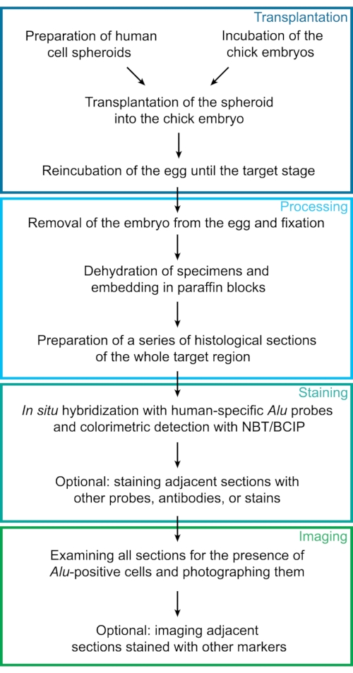
Figure 1: Experimental workflow. This diagram represents the experimental steps of the protocol described here. Individual steps were sorted into 4 major groups: transplantation, processing, staining, and imaging. Abbreviations: NBT = 4-Nitro blue tetrazolium chloride; BCIP = 5-bromo-4-chloro-3-indolyl phosphate p-toluidine. Please click here to view a larger version of this figure.
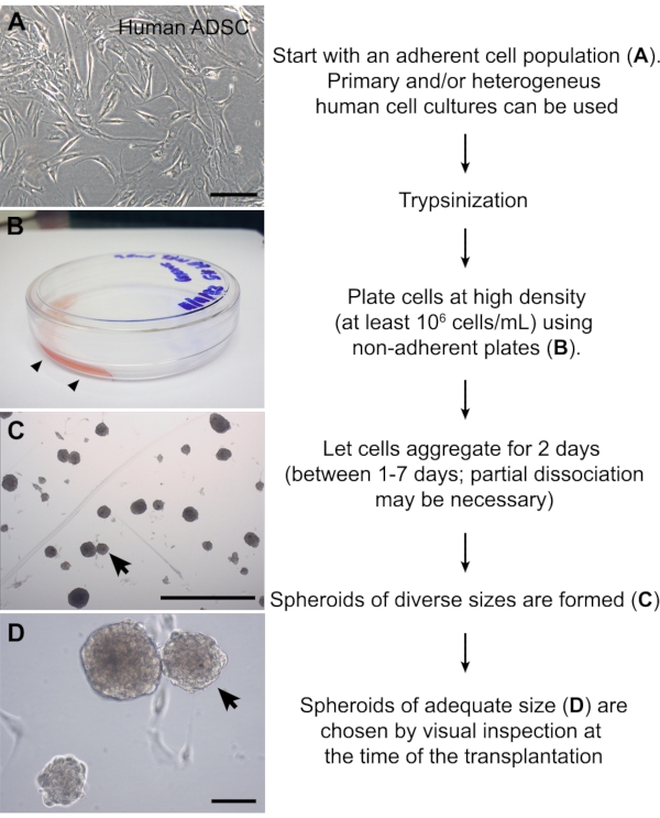
Figure 2: Preparation of cell spheroids. (A) Image of a human adipose-derived stromal cell culture at passage #6. (B) Non-adherent plate in which a small volume (1 mL) of cell suspension was carefully added to one side of the plate (arrowheads). The high cellular density promotes aggregation of the adherent cells. (C, D) ADSC spheroids 2 days after partial dissociation. This method yields spheroids of diverse sizes. A spheroid with the approximate size of the chick somite (~150 µm) is indicated (arrows). Scale bars = 100 µm (A, D) and 1 mm (C). Abbreviation: ADSC = adipose-derived stromal cell. Please click here to view a larger version of this figure.

Figure 3: Transplantation of the spheroid into the presomitic mesoderm of 2-day-old chick embryos. (A) Image of the workspace. Left side: stereomicroscope and gooseneck lamp. Right side: bottle of sterile PBS, India ink, dish with India ink solution in PBS, wide adhesive tape, a pair of microforceps, surgical scissors, slotted spoon, iris scissors, needle holder, egg holder, and Pasteur pipette. Surgical materials have been previously sterilized. (B) Thick capillary for transferring spheroids (left), thin capillary for injecting India ink (middle), and tip of the needle holder with an attached needle (right). (C) Zoomed-in image of the sharpened needle or microscalpel. (D) Illustration of a top view indicating the regions for cutting open the egg and for aspirating the albumen. (E) Illustration of a top view of an opened egg. If the egg has not been rotated in 24 h, the embryo will always sit on the top of the yolk. India ink should be injected into the yolk under the embryo. (F) Photograph of a 2-day-old chick embryo after injection of India ink. (G, H) A chick embryo with 14 somite pairs prior to the grafting of a spheroid, which had already been deposited next to the embryo (arrow). The last formed somite pair (14 s.) is indicated. A cut has been made (arrowhead) in the presomitic mesoderm of the presumptive 16th-17th somites into which the spheroid will be transplanted. (I, J) The same embryo after the transplantation is complete. Abbreviation: PBS = phosphate-buffered saline. Please click here to view a larger version of this figure.
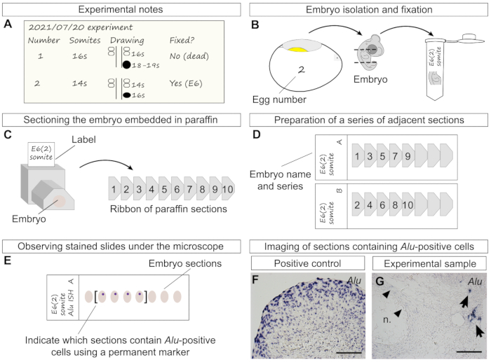
Figure 4: Additional experimental details from transplantation to staining. (A) Information that should be noted throughout the transplantation, including individual egg number, somite stage, and simple drawing of the spheroid position and size in relation to the two last formed somite pairs and neural tube. Later, the stage at which the embryo has been fixed should also be noted. Embryo "2" represents the transplantation performed in Figure 3G–J. (B) Representation of the removal of embryo "2" from the egg at age 6 days, decapitation, removal of the lower half of the trunk, and final transfer of the specimen to a tube containing fixative. (C) Left side: drawing of a trimmed and labeled paraffin block with embedded specimen in transverse orientation. Right side: a ribbon of sequential sections (right side) is obtained from cutting the paraffin block with the microtome. Numbers represent the order in which sections were cut, in an anterior-posterior order. (D) Example of slides prepared as a series of two adjacent sections (series "A" and "B"). The previously cut sections (C) were distributed between two slides, allowing the researcher to perform more than one type of staining of the same sample. (E) Human cells are stained purple-blue after in situ hybridization of Alu probes. Examining the slides and marking sections containing stained cells using a marker pen is recommended, especially sections of older embryos that may span several slides. (F) A large ADSC spheroid was processed for paraffin sectioning and in situ hybridization as positive control. All cells were stained with Alu probes. (G) In this experimental sample (6-day-old chick embryo), human cells were found next to the notochord after hybridization with Alu probes. Chicken cells were not stained. As light background staining may sometimes be found (arrowheads), it is recommended to use a counterstain. Scale bars = 100 µm (F, G). Abbreviation = n. = notochord. Please click here to view a larger version of this figure.
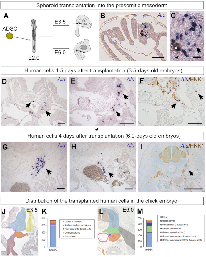
Figure 5: Identification of human ADSC spheroids grafted into the chick presomitic mesoderm and their behavior. (A) Illustration of the transplantation of an ADSC spheroid into the chick presomitic mesoderm. Stages in which embryos were fixed are also indicated, as well as sectioning planes (dashed lines). (B, C) When embryos were fixed only 4 h after the transplantation, the spheroid of human cells still had clearly defined borders29. Human nuclei were stained with Alu probes (arrows), while chicken nuclei were not stained (arrowheads). India ink inclusions were occasionally found next to the implantation site (asterisk). (D–F) When embryos were fixed 1.5 days after the transplantation, the human ADSCs (arrows) did not remain organized as a spheroid. Some cells were found lateral to the neural tube in the sclerotome, while others had migrated ventrally. Co-staining with Alu probes and HNK1, an antibody that recognizes neural crest cells and nerves, revealed a close association between some of the grafted human cells and migratory neural crest cells29. (G–I) When embryos were fixed 4 days after the transplantation, human ADSCs were scattered from the region lateral to the neural tube and notochord (G) to the region surrounding the dorsal aorta (H); many of the latter were perivascular cells. Co-staining with HNK1 revealed many human ADSC cells associated with the peripheral nervous system of the chick29 (I). (J–M) It is possible to classify the grafted human cells according to their distribution, revealing their possible migration from the graft site. Embryos from Cordeiro et al.29; graphs published here (K, 750 Alu-positive cells found in 4 E3.5 embryos; M, 945 Alu-positive cells found in 6 E6.0 embryos). Scale bars = 100 µm (B, D–H), 20 µm (C), and 500 µm (I). Abbreviations: hADSC = human adipose-derived stromal cell; HNK1 = Human Natural Killer 1/ CD57. Please click here to view a larger version of this figure.
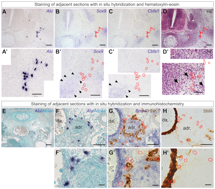
Figure 6: Staining adjacent sections of the same series clarifies the behavior of the grafted human cells. (A, A') Human ADSCs grafted into the chick embryo were located in an E6.0 embryo using Alu probes. (B–D) In situ hybridization of the adjacent sections with the chondrogenic marker Sox9 (B) or the osteogenic marker Cbfa1 (C), as well as observation of the tissue morphology with hematoxylin-eosin (D). (A'–D') Higher-magnification images of panels (A–D). Human cells (red outlines) were not located in skeletogenic territories of the chick vertebra (black arrowheads)29. Nerve fibers (black arrows) can be identified by their morphology with HE staining. (E, F) Human ADSCs were found in the aorta-gonad-mesonephros region of E6.0 embryos. (G). Staining of an adjacent section with Bmp4 probes and HNK1 antibody revealed that human cells (red outlines) were associated with the adrenal gland primordia of the chick. (H). Some cells had a perivascular location (smooth muscle actin)29. (F'–H') Higher-magnification images of panels (F–H). Scale bars = 100 µm (A–E), 50 µm (A'–D', F–H), and 10 µm (F'–H'). Abbreviations: ADSCs = adipose-derived stromal cells; HE = hematoxylin-eosin; da = dorsal aorta; adr = adrenal gland primordia; SMA = alpha smooth muscle actin ; HNK1 = Human Natural Killer 1/ CD57. Please click here to view a larger version of this figure.
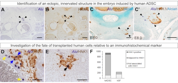
Figure 7: Additional experiments that can give insights into the behavior of grafted human cells. (A–C') In some embryos, an ectopic structure formed by human ADSCs (stained with Alu probes), chick mesenchyme, and chick HNK1-positive peripheral nerves was found (arrowheads). This supports the paracrine effect of ADSCs over their environment. (A, B) n = 2/6 E6 embryos. (C, C') n = 1/2 E8 embryo. (D–F) Quantification of grafted human ADSCs (945 cells, n = 6 embryos29) or skin fibroblasts in relation to HNK1-positive tissue in E6.0 embryos (235 cells, n = 3 embryos29). Alu-positive cells were classified as stained with HNK1 (black arrow), adjacent to HNK1-positive tissue (blue arrows), or neither stained nor adjacent to HNK1-positive tissue (yellow arrows). Scale bars = 100 µm (A–C, C') and 50 µm (D, E). Abbreviations: hADSC = human adipose-derived stromal cell; hSF = human skin fibroblast; HNK1 = Human Natural Killer 1/ CD57. Please click here to view a larger version of this figure.
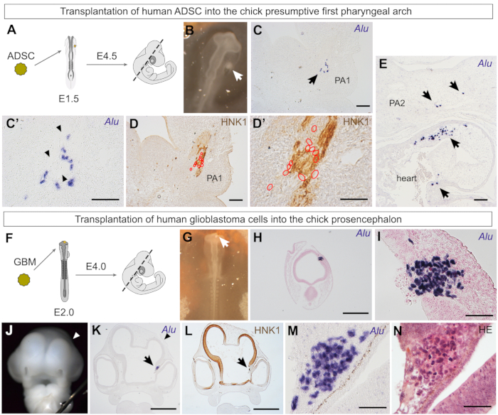
Figure 8: Investigation of the behavior of human ADSCs and glioblastoma cells grafted in the cephalic region of chick embryos. (A) Illustration of the transplantation of an ADSC spheroid into the chick presumptive first pharyngeal arch region, fixation stage, and sectioning plane (dashed line). (B) Human ADSC spheroid (white arrow) grafted between the ectoderm and endoderm, toward which neural crest cells that form the first pharyngeal arch will migrate. (C) Alu-positive cells (arrow) were found in the mandibular bud, a first pharyngeal arch derivative (PA1). (C') Higher magnification revealed that human cells were associated with a structure with scant nuclei (black arrowheads). (D) Human cells (red outlines) were associated with an HNK1-positive peripheral nerve. (D') As seen in a higher magnification image, most human cells (red outlines) are HNK1-negative. (E) Human ADSCs (arrows) were also found in the outflow tract, a region with contribution of cardiac neural crest cells in the chick heart. PA2, second pharyngeal arch. (F) Illustration of the transplantation of a glioblastoma spheroid into the chick prosencephalon, fixation stage, and sectioning plane (dashed line). (G) Photograph of a spheroid of primary human glioblastoma cells grafted into the chick prosencephalic wall. (H, I) Four hours after implantation, human glioblastoma cells were found embedded into the wall of the chick neural tube. Alu-positive cells did not appear integrated into the neuroepithelium at this stage. (J) Photograph of a 4-day-old chick embryo, 2 days after implantation of glioblastoma cells. The right telencephalic vesicle (arrowhead) was smaller than the left (control) side in all embryos (n = 4). (K) Grafted glioblastoma cells (arrow) were found in the diencephalon next to the eye; the reduced telencephalon was also evident in histological sections (arrowhead). (L) HNK1 staining revealed that the neuroepithelium formation was disturbed by the grafted glioblastoma cells. (M, N) Higher magnification of Alu-positive glioblastoma cells (M), clearly discernible from chick cells after hematoxylin-eosin staining (N). Scale bars = 100 µm (C–E), 500 µm (H, K, L), and 50 µm (C', D',I, M, N). Abbreviations: ADSC = adipose-derived stromal cells; GBM = glioblastoma; HNK1 = Human Natural Killer 1/ CD57; HE = hematoxylin-eosin. Please click here to view a larger version of this figure.
Table 1: DNA in situ hybridization solution recipes. Abbreviations: SSC = saline sodium citrate; DEPC = diethyl pyrocarbonate; MAB = maleic acid buffer; NTM = NaCl-Tris-MgCl2; 4-Nitro blue tetrazolium chloride; BCIP = 5-bromo-4-chloro-3-indolyl phosphate p-toluidine. Please click here to download this Table.
Discussion
The protocol described here (Figure 1) presents a feasible option for screening the behavior of primary populations of human cells in vivo, using chick embryos as a model. This paper describes the formation of cell spheroids (Figure 2), transplantation of the spheroid into the chick embryo (Figure 3), processing of specimens and in situ hybridization (Figure 4), representative results of human ADSCs grafted into the presomitic mesoderm (Figure 5) and associated experiments (Figures 6 and 7), as well as results of human ADSCs grafted into the first pharyngeal arch region and human primary glioblastoma grafted into the chick prosencephalon (Figure 8).
This protocol is well adapted for the use of heterogeneous adherent populations, including diverse stromal/mesenchymal cells or tumors, as the label-free detection method eliminates issues with transfection efficiency or selection of specific subpopulations. This is possible due to the smaller scale of the chick embryo and the diverse cell fates maps already established for this organism. The use of spheroids allows human cells to be transplanted into a precise location and interpretation of cell behavior in response to its surrounding microenvironment. Thus, the experimental design is crucial for interpreting the behavior of human cells as changes in the embryonic region or stage will expose grafted cells to a different microenvironment. Regardless of the grafted region, embryos must be reincubated for only a few days to obtain preliminary data on possible migration of grafted cells before investigating a well-defined target region of older embryos.
As done with chick embryo experiments in general, incubating an additional 10% of eggs compensates for unfertilized eggs and loss of viability during transportation from the hatchery. In addition, some embryos die after surgical manipulation and not specifically by the transplantation of cell spheroids. Approximately 10% of the embryos die until E3.5, and up to 40% die until E6.0. Taking care not to damage the yolk membrane or blood vessels during the experiment increases the survivability of the embryos. Good experimental practices during the surgery, including sterilizing surgical materials and adding enough PBS to prevent the embryo from drying out, can also improve the survival rate of the embryos.
The optimal aggregation time and the need for partial dissociation of spheroids depend on the cell type and should be determined empirically. Based on experience, cell lineages and stromal cells tend to form spheroids faster. Moreover, cells that produce an abundant extracellular matrix tend to adhere to the bottom of the plate or form a single, large aggregate. Large spheroids should be partially dissociated by gentle pipetting and then left undisturbed for at least another day To counter these issues. Other cell types may require incubation for shorter or longer times. For example, quail fibrosarcoma cells (QT6) form spheroids after 1 day and do not require dissociation32. In contrast, primary human glioblastoma cells should be gently dissociated after 2-3 days, when 1 mL of additional culture medium can be added and then incubated for a total of 7 days. Alternative methods for preparing cell spheroids may be employed, such as the hanging-drop method33 or agarose-coated wells34. These methods yield individual spheroids with a consistent size according to the number of plated cells. In this case, the experimenter should determine the number of cells required for preparing a spheroid with optimal size.
As human cells are not labeled, it is important to ensure the quality of the histological sections; in situ hybridization procedures enable the location of the grafted cells in the specimens. Important steps include allowing for sufficient time during dehydration to preserve tissue integrity and carefully handling the slides during staining procedures. It is critical to use a positive control in all experiments, especially after the synthesis of new probes. A PCR-cleaning kit may be used to purify the probes, although it was not necessary here. Alu DNA probes exclusively stain human nuclei; cytoplasmatic staining may indicate phagocytosis of human DNA by host cells, appearing as round vesicles inside a larger cell45. Endogenous alkaline phosphatase may sometimes cause cytoplasmatic staining, which can be blocked with levamisole treatment. Immunohistochemical methods for identifying human cells exist and can be alternatively used, albeit they are more costly to use on a large scale. These include, for example, antibodies that detect human nuclear (Ku80, human nuclear antigen, and Lamin B1) and nucleolar antigens (NM95). In contrast, targeting species-specific short interspersed elements (SINEs, such as Alu in humans) can be adapted to identify xenografts in other mammalian species40,64.
Humanized or SCID mice are also widely used xenograft models65,66 that offer an adult microenvironment for grafted cells and thus may answer different scientific questions compared to embryonic xenografts. In addition, chick embryos at early developmental stages are more affordable and present fewer ethical issues as an initial screening model than adult mice. The model presented here is also distinct from the chicken chorioallantoic membrane (CAM) assay, which is performed in the extraembryonic membranes of older embryos and usually aims to investigate the interaction of cells or effects of pharmacological agents on the vasculature3. Furthermore, despite the many useful applications of zebrafish as a xenograft model, the use of a non-amniote model presents intrinsic differences, such as temperature metabolism and oxygen consumption67 that may affect cell behavior, including major events such as heart and fin regeneration capacity68,69. Nevertheless, some limitations are present in the chick embryo xenograft model. It cannot be automated and requires some expertise to perform the graft. By design, it is a small-scale model because of which not many cells can be grafted at once in spheroid form. It is important to avoid using spheroids that are too large as they may have hypoxic centers70,71. If a greater number of cells are required to answer a specific scientific question, more than one spheroid should be grafted instead. In addition, if the transplanted cells are not adherent or if the target region is a cavity or vessel (e.g., neural tube, coelom, or blood vessel), injection of a cell suspension should be performed instead11. In conclusion, the chick embryo is a versatile model that can provide invaluable insights into the behavior of transplanted human cells.
Disclosures
The authors have nothing to disclose.
Acknowledgements
This work was supported by Universidade Federal de Rio de Janeiro (UFRJ for J.B.), Conselho Nacional de Desenvolvimento Científico e Tecnológico (CNPq for J.B.) and Fundação Carlos Chagas Filho de Amparo à Pesquisa do Estado do Rio de Janeiro (FAPERJ for J.B.). We thank T. Jaffredo (CNRS, Paris, France) for the Runx2 (Cbfa1) probe. The HNK1 antibody was obtained from the Developmental Studies Hybridoma Bank developed under the auspices of the NICHD and maintained by The University of Iowa, Department of Biological Sciences, Iowa City, IA 52242 USA. We thank V. Moura-Neto for granting access to the microtome and R. Lent for granting access to the microscope. We thank E. Steck for the help in synthesizing Alu probes.
Materials
| Animals | |||
| Gallus gallus eggs | Granja Tolomei | SPF-free | White leghorn chicken |
| Reagents | |||
| Alcian Blue 8GX | Sigma-aldrich | A5268 | |
| AluFw primers | Sigma-aldrich | OLIGO | 5’-CGA GGC GGG TGG ATC ATG AGG T-3’ |
| AluRev primers | Sigma-aldrich | OLIGO | 5’-TTT TTT GAG ACG GAG TCT CGC-3’ |
| Aluminum sulphate | Sigma-aldrich | 368458 | For Nuclear fast red solution preparation |
| Anti-Digoxigenin-AP, Fab fragments | Roche | 11093274910 | Antibody Registry ID: AB_514497 |
| Anti-Human Natural Killer 1 antibody (HNK1, CD57) | Developmental Studies Hybridoma Bank | 3H5 | Antibody Registry ID: AB_2314644 |
| Anti-mouse, goat IgM-HRP | Santa Cruz Biotechnology | sc-2973 | Antibody Registry ID: AB_650513 |
| Anti-mouse, goat IgG (H+L)-HRP | Novex | G-21040 | Antibody Registry ID: AB_2536527 |
| Anti-Smooth Muscle Actin/ACTA2 antibody | Dako | M085129 | Antibody Registry ID: AB_2811108 |
| Aquatex | Merck | 1085620050 | Aqueous mounting agent |
| 5-Bromo-4-chloro-3-indolyl phosphate p-toluidine salt (BCIP) | Sigma-aldrich | B8503-100MG | |
| Blocking Reagent | Roche | 11096176001 | |
| Citric acid | VETEC | 238 | For SSC buffer preparation |
| Collagenase type IA | Sigma-aldrich | SCR103 | |
| dCTP, dGTP, dATP, dTTP set | Roche | 11969064001 | |
| Denhardt solution 50X | Invitrogen | 750018 | For hybridization buffer preparation |
| Dextran sulphate sodium salt | Thermo Scientific | 15885118 | For hybridization buffer preparation |
| DIG RNA Labeling Mix | Roche | 11277073910 | Contains Dig-11-dUTP |
| DMEM low-glucose | Sigma-aldrich | D5523 | |
| 3,3′-Diaminobenzidine tetrahydrochloride (DAB) | Sigma-aldrich | D5905-50TAB | |
| N,N-Dimethylformamide (DMF) | Sigma-aldrich | 227056 | For NBT and BCIP solution preparation |
| Ethylenediaminetetraacetic acid (EDTA) | Sigma-aldrich | E6758 | For trypsin solution preparation |
| Entellan new | Merck | 107961 | Non-aqueous mounting medium |
| Ethanol | Proquímios | N/A | |
| Fetal bovine serum | ThermoFisher | 12657029 | Inactivate at 56 °C before use |
| Formaldehyde 37% solution | Proquímios | N/A | |
| Formamide | Vetec | V900064 | |
| Glacial acetic acid | Proquímios | N/A | |
| India ink | Pelikan | 221143 | |
| L-glutamine solution (200 mM) | Gibco | 25030-149 | |
| Magnesium chloride | Merck | 8147330100 | For NTM buffer preparation |
| Maleic acid | Sigma-aldrich | M0375-500G | For MAB buffer preparation |
| Methanol | Proquímios | ||
| Normal Goat Serum | Sigma-aldrich | NS02L | Inactivate at 56 °C before use |
| 4-Nitro blue tetrazolium chloride (NBT) | Roche | 11585029001 | |
| Nuclear fast red | Sigma-aldrich | 60700 | |
| Paraplast Plus | Sigma-aldrich | P3558 | |
| Penicillin G sodium salt | Sigma-aldrich | P3032 | |
| Phosphate buffered saline (PBS) | Sigma-aldrich | P3813 | |
| Phosphomolybdic acid | Merck | 100532 | |
| Proteinase K | Gibco BRL | 25530-015 | |
| Salmon sperm DNA | Invitrogen | 15632011 | For hybridization buffer preparation |
| Sodium chloride | Sigma-aldrich | S9888 | For SSC, MAB and NTM buffer preparation |
| Streptomycin Sulfate | Sigma-aldrich | S6501 | |
| Taq Polymerase kit | Cenbiot Enzimas | N/A | |
| Tris-HCl | Sigma-aldrich | T5941 | |
| Trypsin | Sigma-aldrich | T4799 | |
| Tween 20 | Sigma-aldrich | P1379 | |
| Xylene | Proquímios | N/A | |
| Microscope and equipments | |||
| Axioplan upright microscope | Carl Zeiss Microscopy | N/A | |
| Axiovision software | Carl Zeiss Microscopy | N/A | |
| Cell incubator | ThermoForma | 3110 | |
| Egg incubator- 50 eggs | GP | ||
| Gooseneck lamp | Biocam | N/A | For egg manipulation |
| Fiji software; Cell Counter plugin | ImageJ | https://imagej.net/software/fiji/ | |
| Laminar flow hood | TROX | 1385 | |
| Nanodrop Lite | Thermo Scientific | ND-LITE-PR | |
| Rotary microtome | Leica Biosystems | RM2125 RTS | For sectioning |
| Stereomicroscope | Labomed | Luxeo 4D | For egg manipulation |
| Sterilization oven | REALIS | 7261690 | For sterelization of surgical materials |
| Consumables | |||
| 0.2 mL (PCR) polypropylene centrifuge tubes | Eppendorf | 30124707 | |
| 15 mL polypropylene conical centrifuge tubes | Corning | CLS430791 | |
| 1.5 mL polypropylene centrifuge tubes | Axygen | MCT-150-C | |
| 2 mL polypropylene centrifuge tubes | Axygen | MCT-200-C | |
| 50 mL polypropylene conical centrifuge tubes | Corning | CLS430829 | |
| Barrier (Filter) Tips, 200 μL size | Invitrogen | AM12655 | For egg manipulation |
| Excavated Glass Block (Staining Block) with Cover Glass | Hecht Karl | 42020010 | |
| Embedding cassettes | Simport | M480 | Used as a paraffin block holder |
| Glass coverslides, 24 x 40 mm | Kasvi | K5-2440 | |
| Glass Pasteur pipettes 230 mm | NORMAX | 5426023 | For preparation of glass capillaries |
| Microtome blades | Leica Biosystems | HIGH-PROFILE-DISPOSABLE-BLADES-818 | For sectioning |
| Parafilm M | Parafilm | P7793 | |
| Plastic Petri dish, 30 mm | Kasvi | K13-0035 | For egg manipulation |
| Plastic Petri dish, 60 mm | Prolab | 0303-8 | For cell spheroids preparation. Should not be treated for cell adhesion. |
| Silanized glass slides (Starfrost) | Knittel Glass | 198 | For sectioning |
| Syringe 1 mL , Needles 26 G (0.45 x 13 mm) | Descarpack | 32972 | For egg manipulation (albumen aspiration) |
| Surgical tools | |||
| Aspirator tube | Drummond | 2-000-000 | For egg manipulation |
| Dissection scissors | Fine Science Tools | 14061-11 | For egg manipulation |
| Microforceps (tweezers) | Fine Science Tools | 00108-11 | For egg manipulation and preparation of glass capillaries |
| Needle holder (adjustable dissection needle chuck) | Fisherbrand | 8955 | For egg manipulation |
| Oil whetstone, 10.000 grit | N/A | N/A | For sharpening needles |
| Pair of small paint brushes | N/A | N/A | For handling paraffin sections. Any brand may be used. |
| Sewing needles | N/A | N/A | For sharpening into microscalpels. Any brand may be used. |
| Sterile disposable scalpel No. 23 | Swann-Norton | 110 | For sectioning |
| Surgical scalpel handle | Swann-Norton | 914 | For sectioning |
| Wecker iris scissors, sharp/sharp | Surtex | SS-641-11 | For egg manipulation |
References
- Herbert, K. E., Lévesque, J. P., Haylock, D. N., Prince, M. The use of experimental murine models to assess novel agents of hematopoietic stem and progenitor cell mobilization. Biology of Blood and Marrow Transplantation. 14 (6), 603-621 (2008).
- Parada-Kusz, M., et al. Generation of mouse-zebrafish hematopoietic tissue chimeric embryos for hematopoiesis and host-pathogen interaction studies. DMM Disease Models and Mechanisms. 11 (11), (2018).
- Lokman, N. A., Elder, A. S. F., Ricciardelli, C., Oehler, M. K. Chick chorioallantoic membrane (CAM) assay as an in vivo model to study the effect of newly identified molecules on ovarian cancer invasion and metastasis. International Journal of Molecular Sciences. 13 (8), 9959-9970 (2012).
- Jung, J. Human tumor xenograft models for preclinical assessment of anticancer drug development. Toxicological Research. 30 (1), 1-5 (2014).
- Ben-David, U., et al. Patient-derived xenografts undergo mouse-specific tumor evolution. Nature Genetics. 49 (11), 1567-1575 (2017).
- le Douarin, N., Kalcheim, C. . The Neural Crest. , (1999).
- Schneider, R. A. Neural crest and the origin of species-specific pattern. Genesis. 56 (6-7), 23219 (2018).
- Santagati, F., Rijli, F. M. Cranial neural crest and the building of the vertebrate head. Nature Reviews Neuroscience. 4 (10), 806-818 (2003).
- Li, L., Clevers, H. Coexistence of quiescent and active adult stem cells in mammals. Science. 327 (5965), 542-545 (2010).
- Douarin, N., et al. le et al. Evidence for a thymus-dependent form of tolerance that is not based on elimination or anergy of reactive T cells. Immunological Reviews. 149 (1), 35-53 (1996).
- Boulland, J. L., Halasi, G., Kasumacic, N., Glover, J. C. Xenotransplantation of human stem cells into the chicken embryo. Journal of Visualized Experiments: JoVE. (41), e2071 (2010).
- Goldstein, R. S. Transplantation of human embryonic stem cells and derivatives to the chick embryo. Methods in Molecular Biology. 584, 367-385 (2010).
- Akhlaghpour, A., et al. Chicken interspecies chimerism unveils human pluripotency. Stem Cell Reports. 16 (1), 39-55 (2021).
- Kulesa, P. M., Morrison, J. A., Bailey, C. M. The neural crest and cancer: A developmental spin on melanoma. Cells Tissues Organs. 198 (1), 12-21 (2013).
- Hendrix, M. J. C., et al. Reprogramming metastatic tumour cells with embryonic microenvironments. Nature Reviews Cancer. 7 (4), 246-255 (2007).
- Singer, N. G., Caplan, A. I. Mesenchymal stem cells: Mechanisms of inflammation. Annual Review of Pathology: Mechanisms of Disease. 6, 457-478 (2011).
- Bianco, P., Robey, P. G., Simmons, P. J. Mesenchymal stem cells: Revisiting history, concepts, and assays. Cell Stem Cell. 2 (4), 313-319 (2008).
- Griffin, M. D., et al. Concise review: Adult mesenchymal stromal cell therapy for inflammatory diseases: How well are we joining the dots. Stem Cells. 31 (10), 2033-2041 (2013).
- Owen, M., Friedenstein, A. J. Stromal stem cells: marrow-derived osteogenic precursors. Ciba Foundation Symposium. 136, 42-60 (1988).
- Bianco, P., Robey, P. G., Saggio, I., Riminucci, M. “Mesenchymal” stem cells in human bone marrow (skeletal stem cells): A critical discussion of their nature, identity, and significance in incurable skeletal disease. Human Gene Therapy. 21 (9), 1057-1066 (2010).
- Zuk, P. A., et al. Multilineage cells from human adipose tissue: Implications for cell-based therapies. Tissue Engineering. 7 (2), 211-228 (2001).
- Bourin, P., et al. Stromal cells from the adipose tissue-derived stromal vascular fraction and culture expanded adipose tissue-derived stromal/stem cells: A joint statement of the International Federation for Adipose Therapeutics and Science (IFATS) and the International Society for Cellular Therapy (ISCT). Cytotherapy. 15 (6), 641-648 (2013).
- Maumus, M., et al. Native human adipose stromal cells: Localization, morphology and phenotype. International Journal of Obesity. 35 (9), 1141-1153 (2011).
- Zannettino, A. C. W., et al. Multipotential human adipose-derived stromal stem cells exhibit a perivascular phenotype in vitro and in vivo. Journal of Cellular Physiology. 214 (2), 413-421 (2008).
- Phinney, D. G. Functional heterogeneity of mesenchymal stem cells: Implications for cell therapy. Journal of Cellular Biochemistry. 113 (9), 2806-2812 (2012).
- Dominici, M., et al. Minimal criteria for defining multipotent mesenchymal stromal cells. The International Society for Cellular Therapy position statement. Cytotherapy. 8 (4), 315-317 (2006).
- Baptista, L. S., et al. marrow and adipose tissue-derived mesenchymal stem cells: How close are they. Journal of Stem Cells. , 73-90 (2007).
- Sowa, Y., et al. Adipose stromal cells contain phenotypically distinct adipogenic progenitors derived from neural crest. PLoS ONE. 8 (12), 84206 (2013).
- Cordeiro, I. R., et al. Chick embryo xenograft model reveals a novel perineural niche for human adipose-derived stromal cells. Biology Open. 4 (9), 1180-1193 (2015).
- Santos, R. d. e. A., et al. Intrinsic angiogenic potential and migration capacity of human mesenchymal stromal cells derived from menstrual blood and bone marrow. International Journal of Molecular Sciences. 21 (24), 1-23 (2020).
- Menezes, A., et al. Live cell imaging supports a key role for histone deacetylase as a molecular target during glioblastoma malignancy downgrade through tumor competence modulation. Journal of Oncology. 2019, 9043675 (2019).
- Brito, J. M., Teillet, M. A., le Douarin, N. M. Induction of mirror-image supernumerary jaws in chicken mandibular mesenchyme by Sonic Hedgehog-producing cells. Development. 135 (13), 2311-2319 (2008).
- Foty, R. A simple hanging drop cell culture protocol for generation of 3D spheroids. Journal of Visualized Experiments JoVE. (51), e2720 (2011).
- de Barros, A. P. D. N., et al. Osteoblasts and bone marrow mesenchymal stromal cells control hematopoietic stem cell migration and proliferation in 3D in vitro model. PLoS One. 5 (2), 9093 (2010).
- Hamburger, V., Hamilton, H. L. A series of normal stages in the development of the chick embryo. Developmental Dynamics. 195 (4), 231-272 (1951).
- Brady, J. A simple technique for making very fine, durable dissecting needles by sharpening tungsten wire electrolytically. Bulletin of the World Health Organization. 32 (1), 143-144 (1965).
- Tickle, C. How the embryo makes a limb: Determination, polarity and identity. Journal of Anatomy. 227 (4), 418-430 (2015).
- Couly, G., Grapin-Botton, A., Coltey, P., le Douarin, N. M. The regeneration of the cephalic neural crest, a problem revisited: The regenerating cells originate from the contralateral or from the anterior and posterior neural fold. Development. 122 (11), 3393-3407 (1996).
- Walker, J. A., et al. Human DNA quantitation using Alu element-based polymerase chain reaction. Analytical Biochemistry. 315 (1), 122-128 (2003).
- Steck, E., Burkhardt, M., Ehrlich, H., Richter, W. Discrimination between cells of murine and human origin in xenotransplants by species specific genomic in situ hybridization. Xenotransplantation. 17 (2), 153-159 (2010).
- Sams, A., Davies, F. M. R. Commercial varieties of nuclear fast red. Stain Technology. 42 (6), 269-276 (1967).
- Lison, L. Alcian blue 8 g with chlorantine fast red 5 B. A technic for selective staining of mucopolysaccharides. Biotechnic and Histochemistry. 29 (3), 131-138 (1954).
- Cordaux, R., Batzer, M. A. The impact of retrotransposons on human genome evolution. Nature Reviews Genetics. 10 (10), 691-703 (2009).
- Brüstle, O., et al. Chimeric brains generated by intraventricular transplantation of fetal human brain cells into embryonic rats. Nature Biotechnology. 16 (11), 1040-1044 (1998).
- Warncke, B., Valtink, M., Weichel, J., Engelmann, K., Schäfer, H. Experimental rat model for therapeutic retinal pigment epithelium transplantation – Unequivocal microscopic identification of human donor cells by in situ hybridisation of human-specific Alu sequences. Virchows Archiv. 444 (1), 74-81 (2004).
- Kasten, P., et al. Ectopic bone formation associated with mesenchymal stem cells in a resorbable calcium deficient hydroxyapatite carrier. Biomaterials. 26 (29), 5879-5889 (2005).
- Baptista, L., et al. Adipose tissue of control and ex-obese patients exhibit differences in blood vessel content and resident mesenchymal stem cell population. Obesity Surgery. 19 (9), 1304-1312 (2009).
- Christ, B., Huang, R., Scaal, M. Formation and differentiation of the avian sclerotome. Anatomy and Embryology. 208 (5), 333-350 (2004).
- Scaal, M., Christ, B. Formation and differentiation of the avian dermomyotome. Anatomy and Embryology. 208 (6), 411-424 (2004).
- Tucker, G. C., Delarue, M., Zada, S., Boucaut, J. C., Thiery, J. P. Expression of the HNK-1/NC-1 epitope in early vertebrate neurogenesis. Cell and Tissue Research. 251 (2), 457-465 (1988).
- Creuzet, S., Schuler, B., Couly, G., le Douarin, N. M. Reciprocal relationships between Fgf8 and neural crest cells in facial and forebrain development. Proceedings of the National Academy of Sciences of the United States of America. 101 (14), 4843-4847 (2004).
- Charrier, J. B., Lapointe, F., le Douarin, N. M., Teillet, M. A. Dual origin of the floor plate in the avian embryo. Development. 129 (20), 4785-4796 (2002).
- Kordes, U., Cheng, Y. C., Scotting, P. J. Sox group e gene expression distinguishes different types and maturational stages of glial cells in developing chick and mouse. Developmental Brain Research. 157 (2), 209-213 (2005).
- Stricker, S., Fundele, R., Vortkamp, A., Mundlos, S. Role of Runx genes in chondrocyte differentiation. Developmental Biology. 245 (1), 95-108 (2002).
- Grottkau, B. E., Lin, Y. Osteogenesis of adipose-derived stem cells. Bone Research. 1 (2), 133 (2013).
- Culling, C. F. A. . Handbook of histopathological and histochemical techniques. Including museum techniques. , (1974).
- Francis, P. H., Richardson, M. K., Brickell, P. M., Tickle, C. Bone morphogenetic proteins and a signalling pathway that controls patterning in the developing chick limb. Development. 120 (1), 209-218 (1994).
- Huber, K., et al. Persistent expression of BMP-4 in embryonic chick adrenal cortical cells and its role in chromaffin cell development. Neural Development. 3, 28 (2008).
- Pouget, C., Gautier, R., Teillet, M. A., Jaffredo, T. Somite-derived cells replace ventral aortic hemangioblasts and provide aortic smooth muscle cells of the trunk. Development. 133 (6), 1013-1022 (2006).
- Kirby, M. L., Hutson, M. R. Factors controlling cardiac neural crest cell migration. Cell Adhesion and Migration. 4 (4), 609-621 (2010).
- Isern, J., et al. The neural crest is a source of mesenchymal stem cells with specialized hematopoietic stem cell niche function. eLife. 3, 03696 (2014).
- Yamazaki, S., et al. Nonmyelinating schwann cells maintain hematopoietic stem cell hibernation in the bone marrow niche. Cell. 147 (5), 1146-1158 (2011).
- Carr, M. J., et al. Mesenchymal precursor cells in adult nerves contribute to mammalian tissue repair and regeneration. Cell Stem Cell. 24 (2), 240-256 (2019).
- Walker, J. A., et al. Quantitative intra-short interspersed element PCR for species-specific DNA identification. Analytical Biochemistry. 316 (2), 259-269 (2003).
- Shultz, L. D., Ishikawa, F., Greiner, D. L. Humanized mice in translational biomedical research. Nature Reviews Immunology. 7 (2), 118-130 (2007).
- Yong, K. S. M., Her, Z., Chen, Q. Humanized mice as unique tools for human-specific studies. Archivum Immunologiae et Therapiae Experimentalis. 66 (4), 245-266 (2018).
- Cordeiro, I. R., Tanaka, M. Environmental oxygen is a key modulator of development and evolution: From molecules to ecology: Oxygen-sensitive pathways pattern the developing organism, linking genetic and environmental components during the evolution of new traits. BioEssays. 42 (9), 2000025 (2020).
- Poss, K. D., Wilson, L. G., Keating, M. T. Heart regeneration in zebrafish. Science. 298 (5601), 2188-2190 (2002).
- Puente, B. N., et al. The oxygen-rich postnatal environment induces cardiomyocyte cell-cycle arrest through DNA damage response. Cell. 157 (3), 565-579 (2014).
- Colton, C. K. Implantable biohybrid artificial organs. Cell Transplantation. 4 (4), 415-436 (1995).
- Meehan, G. R., et al. Developing a xenograft model of human vasculature in the mouse ear pinna. Scientific Reports. 10 (1), 2058 (2020).

.
